
Abstract
Background. Peptides are short chains of amino acids, and their chemical and physical stability limits how long they can be stored and how far a stored sample still matches its label [1, 2, 3]. The published science of peptide degradation is large but scattered across pharmaceutics, protein chemistry, food science and clinical laboratory medicine. Online guidance on peptide storage rarely cites it, and almost never shows the numbers behind a stated storage condition.
Objective. To map the peptide stability literature by degradation pathway and storage condition, to find where the evidence is thin, and to bring the published kinetic values together in one place with their conditions and the arithmetic shown. A further aim was to set those values against the stability conditions defined by the International Council for Harmonisation (ICH) and adopted in Australia by the Therapeutic Goods Administration (TGA) [4, 5].
Methods. A reproducible evidence map, with reporting informed by PRISMA 2020 and its scoping-review extension [6, 7]. PubMed was searched through NCBI E-utilities on 2 November 2026 (Australia/Brisbane) [8]. A base query for peptide stability or degradation was crossed with 12 degradation-pathway term sets and 9 storage-condition term sets, giving a 108-cell matrix, and was then split into five publication periods. Four cells were hand-audited for precision. Kinetic values (half-lives, fold-differences and activation energies) were taken only from primary studies whose abstracts or records report them. Derived values were calculated from the published numbers, and every step is shown. Regulatory conditions were read from the ICH, TGA, World Health Organization (WHO), European Medicines Agency (EMA) and United States Pharmacopeia (USP) documents themselves.
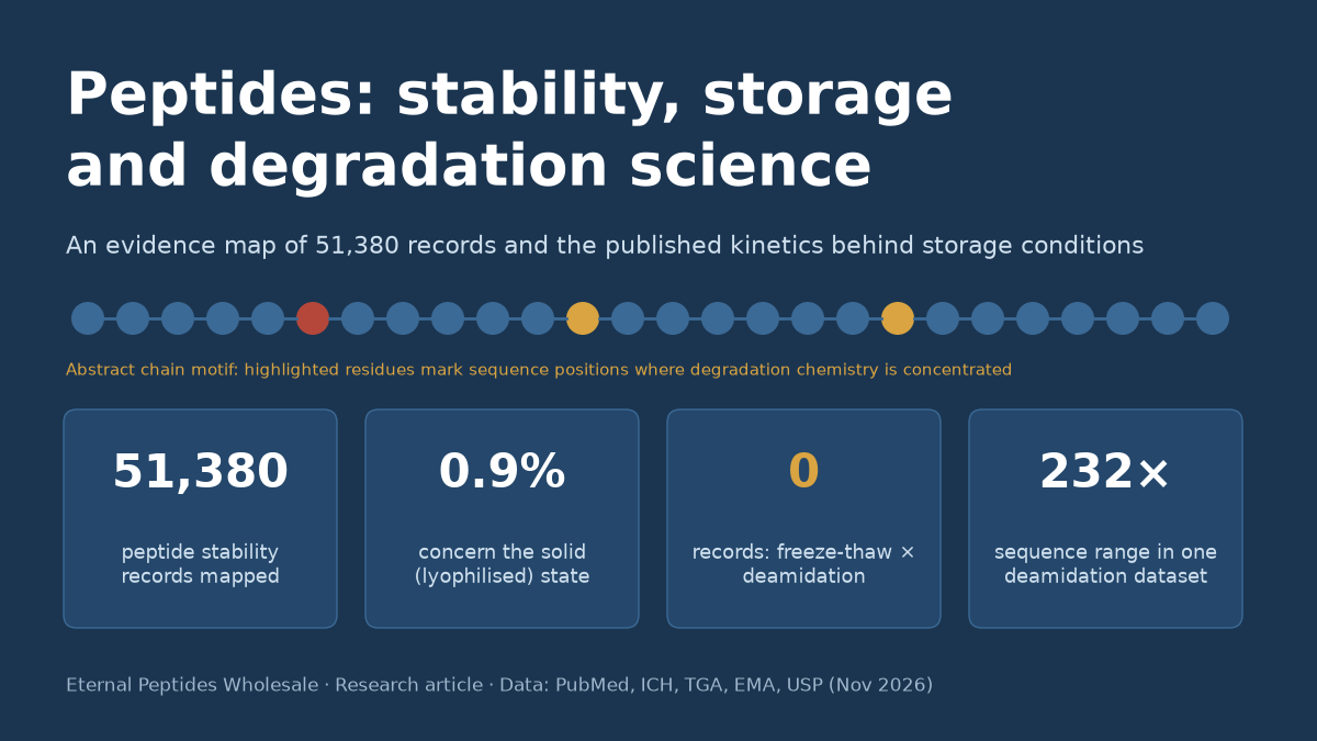
Results. The base query returned 51,380 records. Enzymatic and proteolytic degradation was the largest pathway set (9,346 records; 18.2%). Biological matrices (plasma, serum or blood) were the largest condition set (9,332). Only 476 records (0.93%) concerned the solid or lyophilised state, 210 concerned moisture and 209 concerned freeze-thaw or freezing. Four pathway-condition cells were empty: freeze-thaw crossed with deamidation, isomerization, diketopiperazine formation and pyroglutamate formation. Hand-audit precision was 37 of 56 records (66%). Published half-lives ran from about 4 minutes in diluted serum to model deamidation half-times of up to 50 years, almost seven orders of magnitude. Within a single set of asparagine and aspartate model peptides, sequence alone changed rates 232-fold. Published activation energies of 33.5 to 116.3 kJ/mol imply that cooling from 25 °C to 5 °C slows a reaction 2.6-fold to 29-fold.
Conclusions. Most published peptide “stability” work concerns breakdown by enzymes in biological fluids, not the chemistry of stored material. The storage-relevant evidence is concentrated in a few model hexapeptides and a few proteins. Freeze-thaw chemistry of the peptide bond and side chains is almost unstudied. Temperature effects can be estimated only when an activation energy for the specific reaction is known. Regulatory stability requirements exist because general rules cannot replace product-specific data, and in Australia those requirements assume Zone IV climatic conditions [5].
What is new here. This is, to our knowledge, the first public pathway-by-condition count matrix of the peptide stability literature, with search strings, counts, a precision audit and trend data that anyone can rerun. It brings published degradation half-lives and activation energies into one table with their conditions, derives further values with the arithmetic shown, and uses them to quantify how much a storage temperature changes a degradation rate. It also identifies specific, empty evidence cells where new research is needed.
Key findings
- Of 51,380 PubMed records on peptide stability or degradation, 18.2% concern enzymatic or proteolytic breakdown. Fewer than 1% (476) concern the solid or lyophilised state [8].
- No indexed record combines freeze-thaw with deamidation, isomerization, diketopiperazine or pyroglutamate formation. These are the main non-oxidative chemical routes for stored peptides [8, 9].
- In the classic model hexapeptide, asparagine deamidation has a half-life of 1.4 days at 37 °C and pH 7.4. Replacing the asparagine with aspartate slows the reaction 34-fold [10].
- Neighbouring residues matter as much as the reactive residue itself: one dataset shows a 232-fold range from sequence alone, and pentapeptide models extend deamidation half-times to 2–50 years [11, 12].
- Published activation energies for peptide and lyophile degradation range from about 33.5 to 116.3 kJ/mol. Moving from 25 °C to 5 °C therefore slows degradation 2.6-fold to 29-fold, and a single “rule of thumb” cannot capture that range [13, 14, 15].
- Australia is treated as a Zone IV (hot and humid) region for registration stability studies. A label that says “research use only” does not change whether a peptide product is regulated as a therapeutic good [5, 16].
Contents
- Introduction: why peptides degrade and why the evidence needs mapping
- Peptides degradation chemistry: the pathways
- Methods: searches, counts, audit and kinetic extraction
- Results: the peptide stability evidence map
- Results: how the field has changed since 1966
- Results: published peptide degradation kinetics, with the arithmetic
- Results: what temperature does to peptides degradation rates
- Results: lyophilised peptides, moisture and the solid state
- Results: freezing, freeze-thaw and surface loss
- Results: peptides stability testing rules in Australia and internationally
- Discussion
- Gap analysis: research questions for peptides stability science
- Limitations
- Conclusion
- Relevance to Eternal Peptides Wholesale
- Glossary
- Frequently asked questions about peptides stability and storage
- References
- Related research
- About this article
Introduction: why peptides degrade and why the evidence needs mapping
Peptides are chains of amino acids joined by amide (peptide) bonds. They are usually defined as shorter than proteins, although the boundary is a convention rather than a chemical fact. The TGA’s 2026 safety communication describes them simply as “short chains of amino acids, which are the building blocks of proteins” [1]. More than 80 peptide medicines have reached the market since insulin, and the class sits chemically between small-molecule drugs and biologics [17, 18, 19]. That middle position is the source of most of their stability problems. Like proteins, peptides carry reactive side chains (amides, thiols, thioethers, carboxylic acids and aromatic rings) along a backbone that can be hydrolysed. Unlike most folded proteins, short peptides often have little stable structure to shield those groups, so a reactive residue in a flexible chain is exposed to water, oxygen, light, surfaces and buffer ions [2, 3, 20].
Degradation matters in research for a simple reason. A peptide sample is only useful as an experimental reagent if what is in the tube still matches what the label and the certificate of analysis say. Deamidation adds about one dalton and an extra negative charge. Isomerization to isoaspartate changes neither mass nor composition but alters the backbone. Oxidation of methionine adds 16 daltons. Aggregation and adsorption can remove peptide from solution without creating any new chemical species at all [21, 22, 23]. Each of these changes can alter how a peptide behaves in an assay, and several are invisible to a simple purity measurement unless the analytical method has been shown to separate them. Regulatory guidelines for synthetic peptide medicines now list these pathways explicitly. The EMA guideline on synthetic peptides, adopted in December 2025 and in effect from 1 June 2026, names oxidation, hydrolysis, isomerization, deamidation, cyclic imide (aspartimide) formation, diketopiperazine and pyroglutamic acid formation, beta-elimination, condensation, disulfide cleavage or exchange and acetylation as routes to degradation products, and asks for aggregation to be investigated [9].
The scientific literature on these reactions is old, large and dispersed. Mechanistic work on asparagine and aspartate degradation began with model peptides in the late 1980s [10, 11, 24], formulation science has produced periodic reviews of protein and peptide stability since 1989 [2, 3, 25], and clinical laboratory medicine has its own literature on whether peptide hormones survive sample handling [26, 27]. Most public guidance on peptide storage, by contrast, consists of unreferenced rules about temperatures and months. A reader therefore cannot tell which statements rest on measured kinetics, which are extrapolations, and which are habits.
This article sets out to make that evidence base visible and countable. It does not offer storage instructions, and it makes no claim about the stability of any particular product. Instead it asks four questions. Where in the literature does peptide stability evidence actually sit, by pathway and by condition? Which combinations have barely been studied? What do the published kinetic numbers say when they are placed side by side with their conditions? And how do those numbers relate to the conditions written into the ICH guidelines that the TGA adopts for medicines registered in Australia [4, 28, 5]?
Peptides degradation chemistry: the pathways
Degradation is conventionally divided into chemical instability, in which covalent bonds are made or broken, and physical instability, in which the covalent structure is unchanged but conformation, association state or location changes [2, 21, 22]. The distinction is useful for analysis but not absolute. Covalent changes can promote aggregation, and aggregation can bring reactive groups together. Table 1 summarises the pathways used in this evidence map, with the sequence features and conditions that the primary literature associates with each.
Deamidation and isomerization at asparagine and aspartate
Deamidation of asparagine is the most studied non-enzymatic reaction of peptides. At neutral and alkaline pH, the backbone nitrogen of the following residue attacks the side-chain carbonyl of asparagine to form a five-membered succinimide ring. The ring then hydrolyses to a mixture of aspartate and isoaspartate, and it can also racemize, producing D-residues [10, 29]. In the hexapeptide Val-Tyr-Pro-Asn-Gly-Ala (VYPNGA), a fragment of adrenocorticotropic hormone used as a model in many later studies, Geiger and Clarke measured a deamidation half-life of 1.4 days at 37 °C and pH 7.4. The succinimide intermediate itself hydrolysed with a half-life of 2.3 hours and racemized with a half-life of 19.5 hours [10]. Tyler-Cross and Schirch found that isoaspartate and aspartate products formed in about a 3:1 ratio at neutral and alkaline pH [30]. In acid, the mechanism changes. Asparagine is then hydrolysed directly, without a succinimide, and only the aspartate product forms [31, 32, 30].
Aspartate residues undergo the same ring closure, but more slowly, producing isoaspartate and D-aspartate without any change in mass [10, 11]. Below pH 3, aspartate also promotes hydrolysis of its own peptide bonds. In the Asp-hexapeptide VYPDGA, Oliyai and Borchardt found specific acid-catalysed hydrolysis of the Asp-Gly bond at pH 0.3 to 3, and at pH 4 and 5 isomerization through the cyclic imide to the isoaspartate peptide alongside continued Asp-X hydrolysis [33]. The Asp-Pro bond is a well-known acid-labile site; its unusual cleavage was reported during protein sequencing in 1970 and has since been studied in synthetic epitope peptides [34, 35]. Formulation reviews treat the asparagine and aspartate reactions together because both run through the same cyclic imide intermediate, whose formation is rate-limiting at physiological pH [29].
Oxidation and photodegradation
Methionine, cysteine, histidine, tryptophan and tyrosine are the residues most susceptible to oxidation [36]. Methionine is oxidized to methionine sulfoxide by hydrogen peroxide, hydroxyl radicals, hypochlorite and other oxidants [37]. Oxidation can be driven by trace peroxides in excipients, catalysed by transition metal ions or initiated by light, and its rate depends on pH, temperature and buffer composition [36, 38]. The oxidant matters as well as the residue. In parathyroid hormone (1-34), hydrogen peroxide and t-butyl hydroperoxide oxidized mainly the two methionines, whereas a radical initiator and peroxide plus iron also oxidized tryptophan, and copper led to histidine oxidation [39]. In relaxin, the two surface methionines oxidized independently, at different rates, in line with their solvent exposure [40]. Photodegradation has its own chemistry. Tryptophan, tyrosine, phenylalanine and cystine are the primary photo-oxidation targets [41], and in lyophilised bovine somatotropin near-ultraviolet light attacked disulfide bonds specifically [42].
Diketopiperazine, pyroglutamate and backbone cleavage
A free N-terminal amine can attack the second peptide bond and cut off the first two residues as a cyclic dipeptide, a diketopiperazine. The reaction is fastest when proline is the second residue [43]. In the model compound Phe-Pro-p-nitroanilide, Goolcharran and Borchardt found that the reaction depended on ionization of the N-terminal amine, with a kinetic pKa of 6.1, and on the residue before proline [43]. For substance P, an 11-residue neuropeptide with proline at positions 2 and 4, spontaneous degradation in the solid phase and in solution released the diketopiperazines cyclo(Arg-Pro) and cyclo(Lys-Pro). The acetate salt was less stable than the hydrochloride and trifluoroacetate salts [44]. Later ion-mobility work showed that this sequential cleavage begins with a trans-to-cis change at the penultimate proline [45], and a 2022 mass spectrometry study estimated activation enthalpies of 93 to 109 kJ/mol for diketopiperazine-driven cleavage [46].
N-terminal glutamine, and more slowly glutamate, can cyclize to pyroglutamate, losing ammonia or water. In recombinant antibodies, the half-life of N-terminal glutamate was about 9 months in a pH 4.1 buffer at 45 °C, with minimal conversion at pH 6.2 [47]. The same conversion proceeds in vivo after injection, which shows that it is not only a storage artefact [48]. Ordinary backbone hydrolysis, meanwhile, is slow at neutral pH but is accelerated by acid, by neighbouring aspartate and by specific sequences such as Asn-Ser, where Tyler-Cross and Schirch measured peptide cleavage at about 10% of the rate of deamidation [30].
Aggregation, adsorption and disulfide exchange
Physical instability covers self-association into amorphous aggregates or structured fibrils, adsorption to surfaces, and precipitation [49, 22]. Peptide aggregation depends on intrinsic properties such as sequence, charge and hydrophobicity, and on external factors such as concentration, pH, ionic strength, temperature, interfaces and agitation [49]. Glucagon, a 29-residue peptide hormone, is the standard example. Its fibrils differ in kinetics, morphology and stability depending on salt, peptide concentration and temperature, and pre-formed fibrils pass their properties on when used as seeds [50]. Free thiols and disulfide bonds add another set of reactions, including oxidation, beta-elimination and disulfide exchange, which can drive covalent aggregation [51]. Adsorption is often overlooked because it changes the amount of peptide in solution rather than its structure. It can nevertheless be large: in one systematic study, glass and plastic surfaces removed 90% or more of some cationic peptides from dilute solution [52].
Enzymatic degradation in biological matrices
In plasma, serum, tissue homogenates and cell culture, peptides are cut by proteases and peptidases far faster than by any of the chemical routes above. Powell and colleagues reported half-lives from about 4 minutes to more than 10,000 minutes in diluted human serum, with most peptides at about 10 to 100 minutes [53]. Exopeptidase cleavage at the termini dominated, and placing D-amino acids at both ends stabilized the peptides [54]. Böttger and colleagues found that the same peptides degraded faster in serum than in plasma and were more stable in fresh whole blood, so the choice of matrix itself changes the measured result [55]. Shortening somatostatin and introducing a D-amino acid produced octreotide, whose plasma half-life of about 1.5 hours compares with a few minutes for the parent hormone [56]. This enzymatic literature is important for biology, but it answers a different question from storage stability: it describes what happens to a peptide inside a biological system, not in a sealed vial.
| Pathway | Main sequence or structural features | Conditions that accelerate it in the cited studies | Typical product or change | Key sources |
|---|---|---|---|---|
| Asn deamidation | Asn followed by Gly (fastest), then Ser; bulky or branched residue after Asn slows it up to 70-fold | Neutral to alkaline pH, higher temperature, buffer catalysis; direct hydrolysis in acid | Asp and isoAsp (about 1:3 at neutral pH); +0.98 Da; new negative charge | [10, 30, 31] |
| Asp isomerization | Asp-Gly, Asp-Ser; His next to Asp speeds succinimide formation 8- to 11-fold | pH 4–5 in the model hexapeptide; neighbouring His | isoAsp, D-Asp; no mass change | [33, 57, 58] |
| Acid-catalysed backbone hydrolysis | Asp-X and especially Asp-Pro bonds | pH below about 3 | Fragments | [33, 34, 35] |
| Oxidation | Met, Cys, His, Trp, Tyr | Peroxides, metal ions, light, oxygen | Met sulfoxide (+16 Da), Trp and His oxidation products | [36, 37, 39] |
| Photodegradation | Trp, Tyr, Phe, cystine | UV and visible light; ICH Q1B exposure | Photo-oxidation products, disulfide cleavage | [41, 42, 59] |
| Diketopiperazine formation | Free N-terminus with Pro (or Gly) at position 2 | Unprotonated N-terminal amine (kinetic pKa 6.1 in one model); counter-ion | Cyclic dipeptide plus truncated peptide | [43, 44, 46] |
| Pyroglutamate formation | N-terminal Gln or Glu | pH 4 and pH 8 faster than pH 6.2 for Glu; heat | pyroGlu; −17 Da (Gln) or −18 Da (Glu) | [47, 48] |
| Disulfide exchange and beta-elimination | Free Cys, disulfide bonds | Neutral to alkaline pH, heat | Scrambled disulfides, covalent aggregates | [51] |
| Aggregation and fibrillation | Hydrophobic and amyloidogenic sequences | High concentration, interfaces, agitation, freezing, temperature, salt | Oligomers, fibrils, particles | [49, 50] |
| Adsorption | Cationic and hydrophobic peptides | Low concentration, glass and some plastics | Loss from solution with no new species | [52, 60] |
| Maillard reaction and excipient adducts | Lys, Arg and N-terminal amines | Reducing sugars such as glucose in lyophilised solids | Sugar adducts | [61, 62] |
| Enzymatic degradation | Exposed termini and protease sites | Serum, plasma, tissue homogenates, cell culture | Fragments; half-lives of minutes to hours | [53, 54, 55] |
Methods: searches, counts, audit and kinetic extraction
Search strategy and evidence matrix
The review followed the logic of a scoping review and evidence map: it set out to chart the extent and distribution of a literature rather than to pool effect sizes. Reporting was informed by PRISMA 2020 and the PRISMA extension for scoping reviews [6, 7]. All searches were run against PubMed through the NCBI E-utilities interface (esearch for counts and identifiers, esummary and efetch for records and abstracts) on 2 November 2026 between 04:06 and 04:12 Australia/Brisbane time (AEST, UTC+10) [8].
A base query captured records whose title or abstract mentions peptides together with stability or degradation, or which are indexed under the MeSH heading Drug Stability: (peptide[tiab] OR peptides[tiab]) AND (stability[tiab] OR degradation[tiab] OR "drug stability"[mh]). This base set was crossed with 12 pathway term sets (Table 1) and 9 storage-condition term sets. Each of the 108 pathway-by-condition intersections was counted separately. The full term strings and the total count for each set are given in Table 2. The base set and three condition sets (solid or lyophilised, freeze-thaw and moisture), together with all 12 pathway sets, were then split into five publication-date periods (1966–1985, 1986–1995, 1996–2005, 2006–2015 and 2016–2025) using the PubMed [dp] field. Further counts were made for storage or shelf-life wording, Arrhenius and activation-energy wording, ICH and forced-degradation wording, and publication types (reviews and clinical trials).
Precision audit
Text-word counts measure where words occur, not what papers study. To estimate how far the counts can be trusted, the most recent records in four cells were retrieved and read at title and abstract level: deamidation × solid state (15 records), aggregation × freeze-thaw (15), diketopiperazine × aqueous solution (11, the whole cell) and oxidation × light (15). A record was coded as on-topic if it reported degradation or stability of a peptide or protein product, or a model peptide, under the named condition. The audit was done by a single reviewer, and its result is reported as a simple proportion.
Kinetic data extraction and derived values
Kinetic values were extracted from primary studies identified through the searches and through the reference lists of the main reviews [2, 3, 62, 29]. A value was included only if the PubMed record or abstract states it with its conditions (temperature, pH, matrix or physical state). The included values are half-lives, shelf-life times (T90, the time to 10% loss), fold-differences between sequences or states, and activation energies. Values reported in kcal/mol were converted with 1 kcal = 4.184 kJ. Where a study reported a percentage loss over a time, a first-order rate constant was derived as k = −ln(fraction remaining)/t, and the half-life as ln 2/k. Where a study reported a fold-difference relative to a measured half-life, the derived half-life was obtained by multiplication. Every derived value is labelled as derived, and the arithmetic is shown in Table 4.
Temperature arithmetic
Temperature effects were calculated with the Arrhenius relationship in ratio form, k(T)/k(Tref) = exp[(Ea/R)(1/Tref − 1/T)], with R = 8.314 J/mol/K and temperatures in kelvin. Five activation energies were used: the lower and upper bounds of the range reported for most degradation pathways in lyophiles (8 and 25 kcal/mol, or 33.5 and 104.6 kJ/mol) [13]; the value for VYPNGA deamidation at pH 9 (13.3 kcal/mol, or 55.6 kJ/mol) [15]; the highest value reported for oxytocin, at pH 4.5 (116.3 kJ/mol) [14]; and the default heat of activation of 83.144 kJ/mol used by USP for mean kinetic temperature unless better data are available [63]. Mean kinetic temperatures were calculated with the USP formula [63, 64]. These calculations illustrate how sensitive rates are to temperature. They are not stability predictions.
Regulatory sources
Regulatory statements were taken from the primary documents: ICH Q1A(R2), Q1B and Q5C [4, 59, 28]; the draft consolidated ICH Q1 guideline and the ICH Q1 work plan [65, 66]; WHO Technical Report Series 1010, Annex 10 [67]; USP general chapter 1079.2 [63]; the EMA guideline on synthetic peptides [9]; the TGA’s stability guidance for prescription medicines [5]; the TGA’s 2026 peptide pages [16, 1]; the Therapeutic Goods Act 1989 [68]; and the current Poisons Standard [69]. TGA web pages time out for automated requests from the research host, so they were read in full through a fetching tool, as recorded in the reference ledger.
| Set | Term string (all terms in [tiab] unless stated) | Records in base set | Share of base |
|---|---|---|---|
| Base | (peptide OR peptides) AND (stability OR degradation OR “drug stability”[mh]) | 51,380 | 100% |
| Degradation pathways | |||
| Deamidation | deamidation OR deamidated OR succinimide | 359 | 0.70% |
| Oxidation | oxidation OR oxidized OR oxidised OR “methionine sulfoxide” | 1,517 | 2.95% |
| Hydrolysis or backbone cleavage | hydrolysis OR “peptide bond cleavage” OR “backbone cleavage” | 2,555 | 4.97% |
| Isomerization or racemization | isoaspartate OR isoaspartyl OR isomerization OR isomerisation OR racemization OR racemisation | 391 | 0.76% |
| Diketopiperazine | diketopiperazine OR diketopiperazines | 56 | 0.11% |
| Pyroglutamate | pyroglutamate OR pyroglutamic OR pyroglutamyl | 200 | 0.39% |
| Disulfide exchange | “disulfide scrambling” OR “disulfide exchange” OR “disulfide shuffling” OR “thiol-disulfide” | 66 | 0.13% |
| Aggregation or fibrillation | aggregation OR fibrillation OR fibril OR fibrils | 2,608 | 5.08% |
| Adsorption | adsorption OR “surface adsorption” OR “nonspecific binding” | 579 | 1.13% |
| Maillard or excipient adducts | maillard OR glycation OR “excipient reactivity” | 207 | 0.40% |
| Photodegradation | photodegradation OR photooxidation OR “photo-oxidation” OR photostability | 126 | 0.25% |
| Enzymatic or proteolytic | protease OR proteolysis OR proteolytic OR peptidase | 9,346 | 18.19% |
| Storage and test conditions | |||
| Solid or lyophilised | lyophilized OR lyophilised OR lyophilization OR lyophilisation OR “freeze-dried” OR “solid state” OR “solid-state” | 476 | 0.93% |
| Aqueous solution | “aqueous solution” OR “aqueous solutions” OR “in solution” | 1,527 | 2.97% |
| Temperature or Arrhenius | temperature OR thermal OR arrhenius | 4,848 | 9.44% |
| pH | pH | 4,871 | 9.48% |
| Freeze-thaw or freezing | “freeze-thaw” OR “freeze thaw” OR freezing | 209 | 0.41% |
| Moisture or humidity | moisture OR humidity OR “water content” | 210 | 0.41% |
| Light | light OR photostability OR UV | 2,742 | 5.34% |
| Excipients or formulation | excipient OR excipients OR formulation OR formulations | 2,097 | 4.08% |
| Biological matrix | plasma OR serum OR blood | 9,332 | 18.16% |
| Supplementary counts (each ANDed with the base) | |||
| Storage or shelf life | storage OR “shelf life” OR “shelf-life” | 1,664 | 3.24% |
| Storage AND solid or lyophilised | Storage set AND solid set | 94 | 0.18% |
| Arrhenius | arrhenius | 33 | 0.06% |
| Activation energy or Arrhenius | “activation energy” OR arrhenius | 89 | 0.17% |
| ICH or stress testing | ICH OR “accelerated stability” OR “forced degradation” OR “stress testing” | 129 | 0.25% |
| Reviews | review[pt] | 6,442 | 12.54% |
| Clinical trials | clinical trial[pt] | 233 | 0.45% |
Results: the peptide stability evidence map
Search yield
The base query returned 51,380 PubMed records [8]. Of these, 6,442 (12.5%) were indexed as reviews and 233 (0.45%) as clinical trials. The 12 pathway sets together captured 18,010 record-hits. Because one record can mention several pathways, this sum exceeds the number of distinct records. Enzymatic and proteolytic wording was by far the most common: 9,346 records, or 18.2% of the base set. It was followed by aggregation or fibrillation (2,608; 5.1%), hydrolysis or backbone cleavage (2,555; 5.0%) and oxidation (1,517; 3.0%). The non-oxidative chemical routes that the EMA lists as characteristic of synthetic peptides were each small. Isomerization or racemization had 391 records, deamidation 359, pyroglutamate 200, disulfide exchange 66 and diketopiperazine formation 56 [9]. Diketopiperazine formation, a recognised route to truncated impurities in proline-containing peptides, therefore accounts for about one record in every 900 (51,380 ÷ 56 = 917).
Among the condition sets, biological matrices (plasma, serum or blood; 9,332 records) and pH (4,871) and temperature (4,848) dominated. Formulation or excipient wording appeared in 2,097 records and aqueous-solution wording in 1,527. The three conditions most directly connected with how material is stored before use were the smallest: solid or lyophilised state (476 records; 0.93%), moisture or humidity (210; 0.41%) and freeze-thaw or freezing (209; 0.41%). Storage or shelf-life wording occurred in 1,664 records, but only 94 of these also mentioned the solid or lyophilised state. Only 89 records mentioned an activation energy or Arrhenius analysis, and 129 mentioned ICH, accelerated stability, forced degradation or stress testing.
The pathway-by-condition matrix
Figure 1 and Table 3 show the 108-cell matrix. Three patterns stand out. First, the matrix is dominated by one row and one column. Enzymatic degradation crossed with biological matrix is the largest cell (1,761 records), followed by enzymatic degradation crossed with pH (898) and temperature (678). Second, the solid-state column is thin everywhere. Its largest cell, aggregation, holds 79 records, and the deamidation cell holds 22, even though deamidation in lyophilised solids is one of the most mechanistically studied topics in pharmaceutics [62, 70, 71]. Third, there are holes. Four cells are empty: freeze-thaw crossed with deamidation, isomerization, diketopiperazine formation and pyroglutamate formation. Three more cells are empty in the moisture column (diketopiperazine, pyroglutamate and disulfide exchange), and photodegradation crossed with freeze-thaw is also zero. In all, 8 of 108 cells (7.4%) contain no records, and a further 16 contain 1 to 5 records.
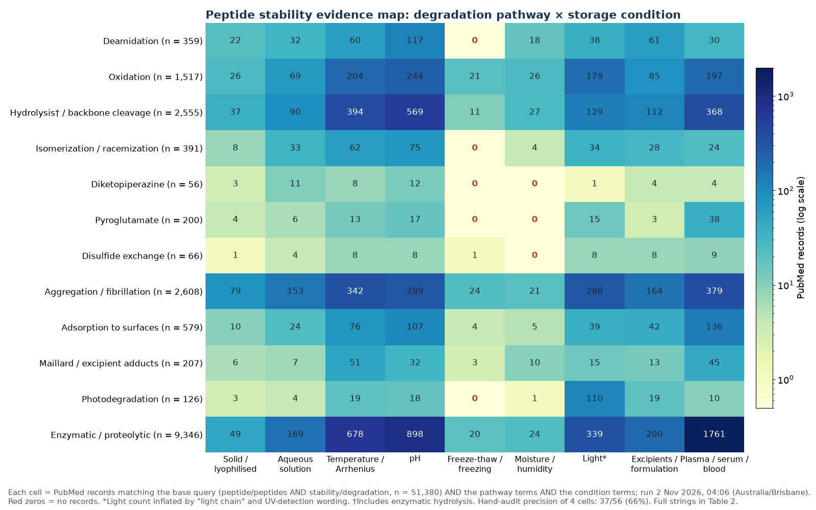
| Pathway | Solid / lyophilised | Aqueous solution | Temperature | pH | Freeze-thaw | Moisture | Light* | Excipients | Plasma / serum / blood |
|---|---|---|---|---|---|---|---|---|---|
| Deamidation | 22 | 32 | 60 | 117 | 0 | 18 | 38 | 61 | 30 |
| Oxidation | 26 | 69 | 204 | 244 | 21 | 26 | 179 | 85 | 197 |
| Hydrolysis / backbone cleavage† | 37 | 90 | 394 | 569 | 11 | 27 | 129 | 112 | 368 |
| Isomerization / racemization | 8 | 33 | 62 | 75 | 0 | 4 | 34 | 28 | 24 |
| Diketopiperazine | 3 | 11 | 8 | 12 | 0 | 0 | 1 | 4 | 4 |
| Pyroglutamate | 4 | 6 | 13 | 17 | 0 | 0 | 15 | 3 | 38 |
| Disulfide exchange | 1 | 4 | 8 | 8 | 1 | 0 | 8 | 8 | 9 |
| Aggregation / fibrillation | 79 | 153 | 342 | 289 | 24 | 21 | 288 | 164 | 379 |
| Adsorption | 10 | 24 | 76 | 107 | 4 | 5 | 39 | 42 | 136 |
| Maillard / excipient adducts | 6 | 7 | 51 | 32 | 3 | 10 | 15 | 13 | 45 |
| Photodegradation | 3 | 4 | 19 | 18 | 0 | 1 | 110 | 19 | 10 |
| Enzymatic / proteolytic | 49 | 169 | 678 | 898 | 20 | 24 | 339 | 200 | 1,761 |
| Precision audit (most recent records, title and abstract): deamidation × solid 15/15 on-topic; aggregation × freeze-thaw 5/15; diketopiperazine × aqueous solution 9/11 (whole cell); oxidation × light 8/15. Total 37/56 (66%). | |||||||||
How far the counts can be trusted
The precision audit gives a sense of how far each cell reflects the topic its label suggests. All 15 of the most recent deamidation × solid-state records were on-topic. They included solid-state insulin kinetics under controlled temperature and humidity [72], a kinetic model of water and deamidation in amorphous solids [70], two mechanism-based studies of succinimide-mediated adducts in lyophiles [73, 74] and three model-peptide studies of sugars, acidic neighbouring residues and beta-turn structure in the solid state [71, 75, 76]. Precision was much lower for aggregation × freeze-thaw (5 of 15). Most off-topic records in that cell came from food science, where freezing of fish, dough or muscle protein is studied for texture and quality rather than for the stability of a peptide. Oxidation × light was 8 of 15 on-topic. The off-topic records used light as a tool (photosensitisers, photoelectrochemical sensors or light-driven bioconjugation) rather than studying it as a stress, and the term “light” can also match “light chain” wording in antibody papers. Diketopiperazine × aqueous solution was 9 of 11. The overall precision of 66% (37 ÷ 56) means the counts should be read as upper bounds on the volume of relevant work, especially for the light and freezing columns. Low precision cannot fill an empty cell, however. A zero is a zero whatever the precision, and the empty cells can only become less empty if relevant work exists that does not use the indexed terms. That possibility is discussed under Limitations.
Results: how the field has changed since 1966
Peptide stability publishing has grown steadily. The base set rose from 2,176 records in 1966–1985 to 20,834 in 2016–2025, a 9.6-fold increase (20,834 ÷ 2,176 = 9.57). Together the five periods hold 48,237 records. The remaining 3,143 base records carry 2026 or other publication dates. Because the whole literature grew, Figure 2 expresses each subset per 1,000 base records in the same period, which separates a change in emphasis from general growth.
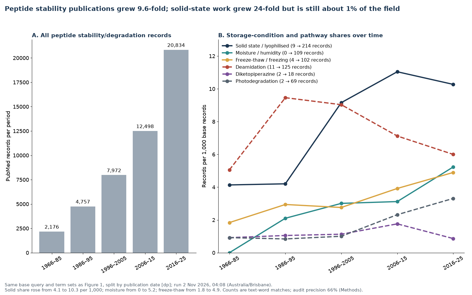
Solid-state work grew 23.8-fold, from 9 to 214 records, faster than the field as a whole. Its share rose from 4.1 to 11.0 per 1,000 base records by 2006–2015 and then eased to 10.3. Even at its peak it was about 1% of the literature. Freeze-thaw work rose from 1.8 to 4.9 per 1,000 and moisture work from zero to 5.2 per 1,000. Aggregation saw the largest change in emphasis, rising from 18.8 to 60.4 per 1,000 (41 to 1,258 records), in line with the attention biopharmaceutical development has given to particles and immunogenicity [49, 25]. Photodegradation rose from 0.9 to 3.3 per 1,000, and Maillard and excipient-adduct wording from zero to 5.1. Enzymatic degradation remained the largest single theme but fell in relative terms, from 246.8 to 150.2 per 1,000.
The classical chemical pathways tell a different story. Deamidation rose from 5.1 per 1,000 in 1966–1985 to 9.5 in 1986–1995, when the model-peptide studies of Clarke, Borchardt and colleagues appeared [10, 11, 31, 77], and has since fallen back to 6.0. Isomerization followed the same arc (3.2, then 10.5, then 6.1). Pyroglutamate fell from 10.6 to 1.6 per 1,000, and diketopiperazine formation stayed at about one record per 1,000 throughout (0.9 to 1.8). In absolute terms, 125 deamidation records and 18 diketopiperazine records appeared in 2016–2025, against 1,258 for aggregation and 3,129 for enzymatic degradation. The mechanistic chemistry of stored peptides is therefore a mature but slow-moving literature, still largely built on studies from the 1980s to 2000s.
Results: published peptide degradation kinetics, with the arithmetic
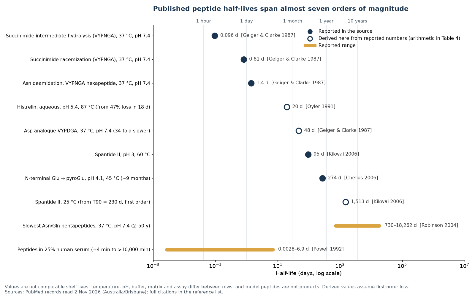
The evidence map shows where the literature is. The kinetic data show what it says. Table 4 brings together half-lives, shelf-life times and fold-differences that are stated with their conditions in primary studies. Figure 3 plots the half-lives on one logarithmic axis. Values in the table fall into three groups: those reported directly, those derived here from reported numbers by the arithmetic shown, and ranges reported as ranges.
| System and pathway | Conditions | Reported value | Derived value and arithmetic | Source |
|---|---|---|---|---|
| VYPNGA hexapeptide, Asn deamidation | 37 °C, pH 7.4 | t½ 1.4 days | Reported | [10] |
| VYPNGA succinimide intermediate, hydrolysis | 37 °C, pH 7.4 | t½ 2.3 hours | 2.3 ÷ 24 = 0.096 days | [10] |
| VYPNGA succinimide intermediate, racemization | 37 °C, pH 7.4 | t½ 19.5 hours | 19.5 ÷ 24 = 0.81 days | [10] |
| VYPDGA (Asp analogue), succinimide formation | 37 °C, pH 7.4 | 34-fold slower than the Asn peptide | 1.4 × 34 = 47.6 days (approximate; assumes the same first-order framework) | [10] |
| Val-Tyr-Pro-X-Y-Ala series, succinimide formation | pH 7.4 | Asn 13.1–35.6× faster than Asp; Gly 6.5–17.6× and Ser 1.6–4.5× faster than Ala at Y; 232-fold overall range | Reported | [11] |
| VSNXV / VXNSV pentapeptides, deamidation | Neutral and alkaline pH | Up to 70-fold slower with bulky or branched residue after Asn, compared with Gly; isoAsp:Asp about 3:1 | Reported | [30] |
| Gly-Xxx-Asn/Gln-Yyy-Gly pentapeptides (648 sequences) | 37 °C, pH 7.4, Tris-HCl | Slowest deamidation half-times 2–50 years | 2 × 365.25 = 730 days; 50 × 365.25 = 18,262 days | [12] |
| His-Asp sequences in ACTH-derived hexapeptides | Physiological conditions | Preceding His increased succinimide formation 8–11-fold | Reported | [58] |
| Histrelin (9-residue analogue), aqueous | 87 °C, pH 5.4 | 47% degraded in 18 days | k = −ln(0.53) ÷ 18 = 0.0353 per day; t½ = 0.693 ÷ 0.0353 = 19.7 days (first-order assumption) | [78] |
| Spantide II, aqueous buffer | 60 °C, pH 3 (maximum stability) | t½ 95 days | Reported | [79] |
| Spantide II, 50% aqueous ethanol | 25 °C | T90 (10% loss) 230 days | k = −ln(0.90) ÷ 230 = 4.58 × 10⁻⁴ per day; t½ = 0.693 ÷ k = 1,513 days (first-order assumption) | [79] |
| N-terminal Glu → pyroGlu (antibody chains) | 45 °C, pH 4.1 | t½ about 9 months | 9 × 30.44 = 274 days | [47] |
| MHC-binding model peptides, enzymatic degradation | 25% human serum or 25% synovial fluid in culture medium | t½ about 4 to more than 10,000 minutes; most 10–100 minutes | 4 ÷ 1,440 = 0.0028 days; 10,000 ÷ 1,440 = 6.9 days | [53] |
| VYPNGA and GQNGG, solid versus solution | 50 °C, pH 7, 5% sucrose or mannitol; solids at 30% RH | 2- to 80-fold slower in solids than in 5% carbohydrate solution | Reported | [71] |
| AcGQNGG and AcGQNQG, solid versus solution | 70 °C; solids at 50% RH with PVP | Up to 60× slower in the solid; acidic N+1 residues showed no solid-state advantage | Reported | [75] |
| Cyclic versus linear beta-turn peptides, solid | 37 °C, PVP lyophiles | Cyclic 1.2–8× slower; rate constants fell by four orders of magnitude in glassy solids | Reported | [76] |
Peptide shelf life: sequence matters as much as the reactive residue
The values span almost seven orders of magnitude, from 0.0028 days (about 4 minutes) for the most labile peptides in diluted human serum to 18,262 days (50 years) for the slowest asparagine and glutamine pentapeptides in buffer. Log10(18,262 ÷ 0.0028) = 6.8. Part of that range reflects the difference between enzymatic and chemical degradation, but a large part comes from sequence alone. In the Val-Tyr-Pro-X-Y-Ala series, changing only the residue at X and the residue after it changed the rate of succinimide formation 232-fold [11]. In the Gly-Xxx-Asn-Yyy-Gly set, the slowest sequences were thousands of times slower than the fastest: the VYPNGA half-life of 1.4 days compares with model half-times of up to 50 years, a ratio of about 13,000 (18,262 ÷ 1.4 = 13,044) [10, 12]. The Robinsons used these rates to build a model that predicts deamidation from primary and three-dimensional structure across 23 proteins [80], and showed by mass spectrometry that synthetic pentapeptides are useful structural models for peptide and protein deamidation [81].
Structure modifies sequence effects further. In folded proteins, native conformation restricts the backbone flexibility that succinimide formation needs, which is why many Asn-Gly sites in proteins are far slower than in the equivalent short peptide [24, 11]. Cyclic beta-turn peptides deamidated 1.2 to 8 times more slowly than their linear analogues in the same solids [76]. Even concentration can matter. In VYPNGA, raising the peptide concentration slowed deamidation, and 5% sucrose reduced the rate by up to 40% at 35 °C, an effect attributed to a greater preference for a beta-turn conformation [15]. Model peptide data can still transfer reasonably well. In one comparison, deamidation and oxidation rates in fragments of a growth factor were comparable to rates in the full protein, although diketopiperazine formation was slower in the protein [82].
pH changes the mechanism, not only the rate
The pH dependence of peptide degradation is not a single curve. Asparagine deamidates through the succinimide at neutral and alkaline pH but by direct side-chain hydrolysis in acid [31, 30]. The size of the residue after asparagine strongly affects the neutral-pH route but not the acid route [32]. Aspartate-containing peptides undergo backbone hydrolysis in strong acid and isomerization at pH 4 to 5 [33], and the size of the residue after aspartate changes how fast the cyclic imide forms [57]. Corticotropin (ACTH) deamidated by pseudo-first-order kinetics that depended on pH and buffer [77]. For whole molecules, the result is a pH of maximum stability that is specific to each peptide. Spantide II was most stable at pH 3 to 5 [79], oxytocin degraded fastest at pH 9.0 and slowest at pH 4.5 [14], and N-terminal glutamate cyclized least at pH 6.2 [47]. Buffer species matter as well. Phosphate and glycine buffers catalysed diketopiperazine formation in a model dipeptide derivative [43], and buffer catalysis of deamidation was seen between pH 7 and 11 [31]. No single “safe” pH exists for peptides as a class.
Results: what temperature does to peptides degradation rates
Temperature is the storage variable that public guidance most often addresses, and the one for which the arithmetic is most often skipped. The Arrhenius equation links rate to temperature through the activation energy (Ea) of the reaction. A review of lyophile stability found that most reported activation energies for degradation pathways in freeze-dried solids fall between 8 and 25 kcal/mol (33.5 to 104.6 kJ/mol). It concluded that the Arrhenius equation is a reasonable empirical tool for these solids “provided that specific conditions are met”, while noting breaks in Arrhenius plots around the glass transition temperature [13]. Deamidation of VYPNGA at pH 9 followed Arrhenius behaviour from 5 to 65 °C with Ea = 13.3 kcal/mol (55.6 kJ/mol) [15]. Oxytocin degradation followed the Arrhenius equation at four pH values, with the highest activation energy, 116.3 kJ/mol, at pH 4.5 [14]. For diketopiperazine-driven cleavage of model Xaa-Pro peptides, activation enthalpies were about 93 to 109 kJ/mol [46].
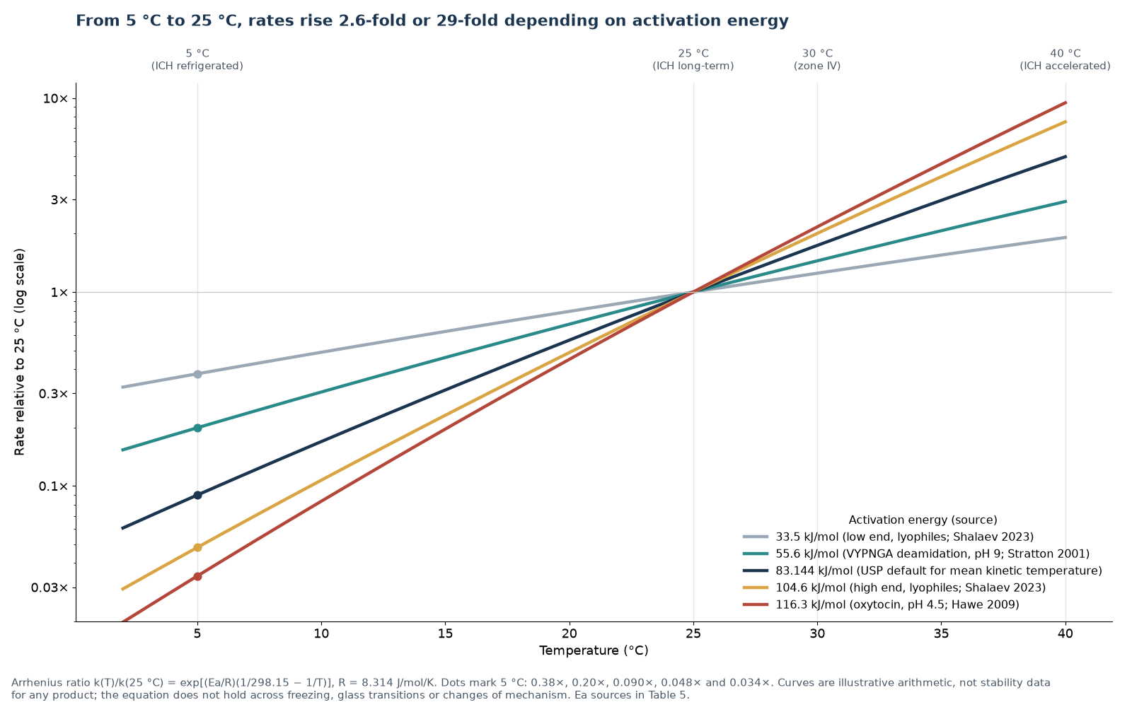
Figure 4 and Table 5 show what these activation energies imply. Using the ratio form of the Arrhenius equation, cooling from 25 °C to 5 °C slows a reaction 2.6-fold at 33.5 kJ/mol, 5.0-fold at 55.6 kJ/mol, 11.2-fold at the USP default of 83.144 kJ/mol, 20.8-fold at 104.6 kJ/mol and 29.2-fold at 116.3 kJ/mol. The worked example for 83.144 kJ/mol is exp[(83,144 ÷ 8.314) × (1/278.15 − 1/298.15)] = exp(10,001 × 0.000241) = exp(2.41) = 11.2. Warming from 25 °C to the ICH accelerated condition of 40 °C speeds a reaction 1.9-fold to 9.5-fold across the same range. The often-quoted rule that a 10 °C rise doubles a reaction rate corresponds, between 20 °C and 30 °C, to an activation energy of about 51 kJ/mol. The five activation energies used here give Q10 values of 1.57 to 4.83, so the rule is right only for some reactions.
The same arithmetic shows why short-term data cannot simply be scaled. As an illustration only, the activation energy measured for VYPNGA at pH 9 can be applied to the half-life at pH 7.4 and 37 °C. That predicts a half-life of 1.4 × 2.38 = 3.3 days at 25 °C and 16.7 days at 5 °C. Extending the same equation to −20 °C gives 180 days, but that number is not valid. Freezing changes phase, concentrates solutes, can shift buffer pH by more than a unit and can create ice-liquid interfaces, so one Arrhenius line cannot describe a reaction across the freezing point [83, 84]. The Arrhenius equation also fails to describe some lyophile reactions across the glass transition [13, 85, 86], and mixed mechanisms, as at different pH values, can give apparently non-Arrhenius behaviour [14]. The ICH guidelines reflect this. Q5C requires that storage periods for biotechnological products be based on “long-term, real-time, real-condition stability studies” [28], and Q1A requires freezer storage periods to be based on real-time data at the long-term condition, with only a single-batch study at an elevated temperature to address excursions [4].
| Quantity | 33.5 kJ/mol | 55.6 kJ/mol | 83.144 kJ/mol | 104.6 kJ/mol | 116.3 kJ/mol |
|---|---|---|---|---|---|
| Source of Ea | Lower bound, most lyophile pathways (8 kcal/mol) | VYPNGA deamidation, pH 9 (13.3 kcal/mol) | USP default for mean kinetic temperature | Upper bound, most lyophile pathways (25 kcal/mol) | Oxytocin, pH 4.5 |
| Rate at 5 °C relative to 25 °C | 0.378 | 0.199 | 0.0897 | 0.0481 | 0.0343 |
| Slow-down from 25 °C to 5 °C | 2.6× | 5.0× | 11.2× | 20.8× | 29.2× |
| Rate at 30 °C (Zone IV long-term) | 1.25× | 1.45× | 1.74× | 2.01× | 2.17× |
| Rate at 40 °C (ICH accelerated) | 1.91× | 2.93× | 4.99× | 7.55× | 9.46× |
| Q10 between 20 and 30 °C | 1.57 | 2.12 | 3.08 | 4.12 | 4.83 |
| Naive slow-down from 25 °C to −20 °C | 11× | 54× | 389× | 1,810× | 4,188× |
| ICH and WHO stability conditions: long-term 25 °C ± 2 °C / 60% RH ± 5% RH or 30 °C ± 2 °C / 65% RH ± 5% RH; intermediate 30 °C / 65% RH; accelerated 40 °C ± 2 °C / 75% RH ± 5% RH; refrigerated long-term 5 °C ± 3 °C with 25 °C / 60% RH accelerated; freezer −20 °C ± 5 °C; Zone IVb long-term 30 °C / 75% RH [4, 67]. | |||||
| Mean kinetic temperature examples (USP formula, ΔH 83.144 kJ/mol): 23 hours at 5 °C and 1 hour at 25 °C give an MKT of 7.8 °C against an arithmetic mean of 5.8 °C; 22 hours at 5 °C and 2 hours at 25 °C give 9.8 °C; 29 days at 25 °C and 1 day at 40 °C give 26.1 °C [63]. | |||||
Mean kinetic temperature (MKT) puts the same point in regulatory form. USP chapter 1079.2 defines MKT as “the single calculated temperature at which the total amount of degradation over a particular period is equal to the sum of the individual degradations”, using a default ΔH of 83.144 kJ/mol “unless more accurate information is available from experimental studies” [63]. Because the calculation is exponential, brief warm periods weigh more than their duration. In the worked examples, one hour at 25 °C in a day otherwise spent at 5 °C raises the MKT to 7.8 °C, although the arithmetic mean is only 5.8 °C. Two hours raise it to 9.8 °C, above the USP limit of 8 °C for controlled cold temperature. USP also states that degradation caused by higher temperatures is not reversible and that MKT must not be used to justify a system that is out of control [63]. The idea of a single “virtual temperature” that integrates a fluctuating temperature history goes back to work by Haynes in 1971 [64].
Results: lyophilised peptides, moisture and the solid state
Lyophilisation (freeze-drying) is the usual way to give peptides and proteins an acceptable shelf life when they are unstable in solution [87, 88]. The evidence map shows that solid-state work is less than 1% of the peptide stability literature, but the work that exists is unusually quantitative. Its central finding is that a dry solid is not a frozen moment. Chemical reactions continue in amorphous solids at rates governed by molecular mobility, residual water and the excipient matrix [62, 89].
The solid state usually slows reactions, by widely varying amounts. VYPNGA and GQNGG deamidated 2 to 80 times more slowly in lyophilised sucrose or mannitol than in 5% solutions of the same carbohydrates, and the relative rates of the two peptides depended on the matrix [71]. AcGQNGG and AcGQNQG were up to 60 times slower in polyvinylpyrrolidone solids than in solution. When glutamate or aspartate followed the asparagine, however, solid-state rates were similar to solution rates, possibly because the acidic residue increases local hydration [75]. Rate constants for model beta-turn peptides fell by four orders of magnitude in glassy solids compared with more mobile matrices [76]. For an aspartate hexapeptide in lyophilised formulations, the type of bulking agent (amorphous or crystalline), residual moisture and temperature were significant variables, while the pH of the starting solution was not [90].
Water affects solid-state reactions in more than one way. Review of the literature shows two patterns for deamidation in amorphous solids: a steady increase in rate with water content, and a “hockey stick” pattern in which deamidation is almost independent of water until a threshold is crossed [70]. In lyophilised insulin, degradation increased as the apparent pH fell, giving solid-state pH-rate profiles parallel to the solution profile, and the reaction plateaued in a way best described by reversible first-order kinetics [91]. Water also changed how the reactive intermediate partitioned between products [92]. In a 2025 study of solid human insulin stored at 25, 40 and 60 °C and 1%, 33% and 75% relative humidity for 6 months, temperature had a major effect on deamidation and covalent aggregation, while humidity mattered little at 25 °C and 40 °C but was important at 60 °C [72]. For a lyophilised antibody, high moisture increased aggregation only when the cake was stored above its glass transition temperature, but increased aspartate isomerization both above and below it. All formulations were stable at 5 °C over 6 to 12 months [85].
Solids create reactions that solutions do not. In amorphous lyophiles of the model peptide Gly-Phe-Asn-Gly, the succinimide formed during deamidation reacted with a neighbouring dipeptide to form covalent amide-linked adducts, a possible route to non-reducible aggregates [73, 74]. Reducing sugars such as glucose formed Maillard adducts with relaxin in lyophilised formulations [61]. In a lyophilised antibody, degradation was slowest at about 2 to 3% water, and adding a little sorbitol to a sucrose formulation improved stability even though it increased molecular mobility [93]. In related work, storage stability correlated best with how much native structure survived drying, and could not be fully explained by glass dynamics [94]. Trehalose, one of the commonest stabilising sugars, has a long record of use in biological preservation [95]. In freeze-dried growth hormone, sucrose and trehalose gave similar chemical stability but different patterns of aggregation [96]. For keratinocyte growth factor-2, the main factor governing degradation was the fraction of protein at the glass solid-air interface [97]. Molecular mobility below the glass transition has been used to explain why aggregation in some sugar glasses is expected to be very slow under refrigeration [86]. Practical formulation advice for lyophilised proteins rests on these mechanisms [98], and early work on deamidation in lyophilised proteins identified this as a solid-state pathway in its own right [99]. Light can also act on solids. Near-ultraviolet light attacked only the disulfide bonds of lyophilised bovine growth hormone, through a tryptophan-mediated, oxygen-dependent mechanism, and residual moisture between 0.4% and 10% did not change the rate [42].
The EMA guideline on synthetic peptides draws practical regulatory conclusions from this literature. It notes that “peptides are often very hygroscopic powders”, says that water content should be part of stability protocols, and states that “Generally, to prevent or minimise degradation, peptides are stored under refrigerated (5°C ± 3°C) or frozen conditions (-20°C ± 5°C)”, with data at higher temperatures and humidities expected to cover short-term excursions [9]. These are regulatory expectations for medicines, supported by product-specific stability data. They are not universal storage rules.
Results: freezing, freeze-thaw and surface loss
Freezing is often treated as the end point of stability, but the protein literature describes it as a stress in its own right. Cooling exposes molecules to low temperature, freeze-concentration and ice surfaces. Freeze-concentration can speed second-order reactions, crystallize buffer components and separate phases [83]. Buffer pH can shift on freezing. Between +25 °C and −30 °C, sodium phosphate buffer showed the largest change of the common buffers, Tris-hydrochloride rose by about 1.2 pH units, and histidine, acetate, citrate and succinate buffers changed by less than one unit [84]. In that antibody study, aggregation after freeze-thaw depended on whether a non-crystallizing cryoprotectant was present rather than on the pH shift [84].
For peptides specifically, the evidence is thin. The audit found only a few peptide-focused freeze-thaw studies among food-science records. Differential scanning calorimetry of glucagon solutions through repeated freeze-thaw cycles showed that lactose stabilised the peptide against aggregation better than trehalose, and that a high excipient-to-peptide ratio reduced aggregation regardless of excipient [100]. A 2026 study of physical stress in liquid peptide solutions found that freezing and thawing in glass vials at −50 °C produced subvisible particles even in peptide-free buffer, possibly from glass delamination, which shows that particle counts after freezing do not necessarily come from the peptide [101]. Clinical laboratory studies, which concern endogenous peptides in plasma rather than isolated material, show mixed results. Across 15 endocrine measures and up to four freeze-thaw cycles, most were unaffected, but plasma renin activity rose and adrenocorticotropic hormone fell slightly [26]. In a 2025 study of 42 analytes, N-terminal pro-brain natriuretic peptide was assessed in serum and plasma after up to four freeze-thaw cycles at −80 °C, and most analytes stayed within bias limits based on biological variation [27]. A review of cytokines found wide variation in reported storage and freeze-thaw stability across 33 cytokines and advised caution [102]. None of these studies measured deamidation, isomerization, diketopiperazine or pyroglutamate formation during freeze-thaw, which is consistent with the empty cells in Figure 1.
Surface loss is the other underestimated route. Cationic membrane-active peptides adsorbed to glass and plastic sample containers so quickly that 90% or more could be lost from solution at typical experimental concentrations [52]. Recovery of radiolabelled endocrine peptides varied widely between glass, plastic and siliconized surfaces. Adding albumin improved recovery, siliconization reduced it, and choosing the most suitable tube for each peptide gave more than 89% recovery after lyophilisation with albumin [60]. These losses involve no chemical change, so a purity assay can still show an apparently pure sample even when much less peptide is present than expected. Agitation, pumping and stirring can likewise generate particles in peptide solutions [101, 49]. Methods that report content or concentration therefore answer a different question from methods that report chromatographic purity [21, 23].
Results: peptides stability testing rules in Australia and internationally
ICH, WHO and USP stability conditions
Formal stability testing for medicines is defined by the ICH Q1 series. Q1A(R2) sets the long-term, intermediate and accelerated conditions summarised in Table 5, and separate conditions for refrigerated and frozen storage [4]. Q1B defines photostability testing, with confirmatory exposure of not less than 1.2 million lux hours of visible light and 200 watt hours per square metre of near-ultraviolet energy [59]. Q5C covers biotechnological products and does not accept accelerated data as a substitute for real-time data [28]. WHO Technical Report Series 1010, Annex 10, applies the same framework to all climatic zones. Its long-term conditions are 25 °C / 60% RH for Zone II, 30 °C / 65% RH for Zone IVa and 30 °C / 75% RH for Zone IVb [67]. USP chapter 1079.2 defines controlled room temperature as storage at 20 to 25 °C with an MKT of not more than 25 °C, and controlled cold temperature as 2 to 8 °C with an MKT of not more than 8 °C, with defined allowances for excursions [63].
The ICH Q1 family is being revised. The Step 2 draft of a consolidated Q1 guideline, dated 11 April 2025, will replace Q1A to Q1F and Q5C. Its scope explicitly includes chemically synthesised polypeptides alongside therapeutic proteins [65]. The ICH work plan dated 10 July 2026 schedules expert consensus in November 2026, Step 3 sign-off in December 2026 and training material in June 2027 [66]. On 2 November 2026, the consolidated guideline was therefore still a draft, and the existing Q1A(R2), Q1B and Q5C remain the adopted texts.
The EMA synthetic peptide guideline
The EMA Guideline on the Development and Manufacture of Synthetic Peptides (EMA/CHMP/CVMP/QWP/367182/2025) was adopted by the CHMP on 1 December 2025 and by the CVMP on 4 December 2025, and came into effect on 1 June 2026 [9]. It notes that synthetic peptides are fully or partly excluded from the scope of ICH Q3A/B, Q6A/B and M7. It lists the degradation pathways summarised above, asks for forced degradation studies to identify likely degradation products, and expects high-molecular-weight impurities and aggregation to be investigated. It also asks for the counter-ion, usually acetate but sometimes trifluoroacetate or chloride, to be defined and controlled. For impurities, the guideline refers to the European Pharmacopoeia thresholds for peptide-related impurities: reporting above 0.1%, identification above 0.5% and qualification above 1.0% [9]. The counter-ion requirement has a research parallel. Trifluoroacetate left over from purification affected bone-cell cultures at 10−8 to 10−7 M, enough to change the apparent result of an assay [103], and the counter-ion also affected the solid-state stability of substance P [44]. Forced degradation itself remains poorly standardised for proteins. Apart from photostability, there are no standard procedures [104].
Australia: Zone IV and therapeutic goods regulation
The TGA adopts the European versions of the ICH stability guidelines with annotations. Its stability guidance for prescription medicines states that major population centres in Australia “experience a combination of high humidity and high temperature during the summer” and are “classified as Zone IV regions”, and that stability studies for medicines to be registered in Australia must be performed under conditions representative of Zone IV [5]. For biological medicines, it describes temperature-cycling studies to support shipping excursions, noting that shipping to Australia “is always lengthy due to the distances involved and crosses the equatorial region” [5]. These requirements show that Australian regulators consider climate and transport duration to be material to stability. They apply to registered medicines and are not a standard for other products.
Stability science says nothing about legal status, and the two should not be confused. Under the Therapeutic Goods Act 1989, peptide products are regulated as therapeutic goods in Australia [1, 68]. The TGA states that disclaimers that a product is for “research use only” or “not for human use” do not change whether a product is regulated as a therapeutic good, and do not remove advertising, manufacture, import, export or supply obligations [16]. Its April 2026 safety alert adds that such a disclaimer “does not make its supply lawful” [1]. Scheduling under the Poisons Standard is specific to each substance and is set out in the current instrument, the Therapeutic Goods (Poisons Standard—October 2026) Instrument 2026 [69]. Nothing in this article about stability, storage or testing implies that any peptide is approved, lawful to supply or suitable for human use.
Discussion: what the peptides stability evidence does and does not support
Principal findings
This evidence map gives the peptide stability literature a measured shape. Three findings stand out. First, the literature is large but concentrated. About 18% of records concern enzymatic degradation and about 18% concern biological matrices, while the solid state, moisture and freeze-thaw conditions that determine how isolated material behaves in storage together account for less than 2%. Second, the chemistry that limits the life of stored peptides is mechanistically well understood but thinly sampled. Deamidation, isomerization, diketopiperazine and pyroglutamate formation together have about a thousand indexed records over six decades, and none of these four pathways has a single record combined with freeze-thaw. Third, the quantitative data show that sequence and activation energy dominate. A change of neighbouring residue can alter a rate 232-fold [11], half-lives in buffer span more than four orders of magnitude [10, 12], and the effect of refrigeration ranges from a 2.6-fold to a 29-fold slow-down depending on the activation energy [13, 14].
Together these findings explain why general statements about peptide shelf life are so often inconsistent. A claim that peptides are “stable for years when frozen” or “stable for weeks in solution” is not wrong for every peptide. It cannot be right for all of them, because the variables that decide stability (sequence, conformation, counter-ion, water content, matrix, pH, temperature history and surface contact) vary independently and have effects of similar size. The literature supports reasoning about a particular molecule in particular conditions. It does not support universal rules.
How these findings compare with earlier reviews
Earlier reviews have catalogued the same chemistry thoroughly. Manning and colleagues reviewed protein stability in 1989, 2010 and again in 2024 [2, 3, 25]. Reubsaet and colleagues reviewed chemical and physical instability of peptides and their analysis [21, 22], Lai and Topp reviewed solid-state chemistry [62], and Zapadka and colleagues reviewed factors that affect peptide aggregation [49]. These reviews describe mechanisms and give examples, but none of them measures how much evidence exists for each pathway and condition. The map adds that measurement. It confirms what narrative reviews imply, that solid-state and freezing chemistry are under-studied relative to their practical importance, and it makes the gaps precise enough to test. The finding that deamidation and isomerization peaked in relative share in 1986–1995 and have declined since is, as far as we know, not reported elsewhere. It suggests that the mechanistic foundation of this field rests on work now 30 or more years old.
The quantitative synthesis also qualifies two common simplifications. The first is the 10 °C rule: as Table 5 shows, Q10 values between 1.6 and 4.8 are consistent with published activation energies, so a doubling per 10 °C describes only some reactions. The second is the assumption that lyophilisation fixes a molecule in place. The solid-state literature shows that deamidation, isomerization, adduct formation and covalent aggregation all continue in dry solids, at rates that depend on water content and matrix [91, 73, 85, 70]. Lyophilisation slows chemistry, sometimes by orders of magnitude, but it does not stop it [76, 71].
Mechanistic interpretation
Most of the patterns in the kinetic data follow from a few mechanisms. Succinimide formation needs the backbone nitrogen of the next residue to reach the side-chain carbonyl, so small, flexible neighbours such as glycine and serine speed it up and bulky or branched neighbours slow it down [11, 30, 10]. Folded structure and cyclization restrict the same motion, which is why proteins and cyclic peptides usually deamidate more slowly than linear model peptides [24, 76]. In solids, the rate-limiting cyclization needs only local flexibility. Bimolecular reactions with water or another molecule depend on wider mobility, which is why water content changes the product mix as well as the rate [92, 91]. Diketopiperazine formation depends on the N-terminal amine being unprotonated and on the residue before proline, giving a sequence- and pH-dependent route that removes two residues at once [43, 46, 45]. Oxidation depends on dissolved oxygen, trace metals, peroxides and light more than on temperature alone [39, 40, 41]. Physical losses through adsorption and aggregation depend on surfaces, interfaces and concentration rather than on covalent chemistry [52, 49, 60].
These mechanisms help explain why a short list of storage rules cannot be universal. A cold, dry, dark, oxygen-poor solid slows most chemical routes. Even so, the extent of protection for any given molecule depends on its sequence, its counter-ion, the excipients present and the surface it touches. Freezing a solution introduces new stresses as well as slowing old ones [83, 84].
Implications for peptide storage research practice
For researchers, the main implication is that stability is an experimental variable, not an assumption. Several practices follow from the evidence. Where a peptide contains Asn-Gly, Asn-Ser, Asp-Gly, an N-terminal Gln or Glu, an Xaa-Pro N-terminus, Met, Cys or Trp, the corresponding degradation products are predictable and can be looked for [11, 47, 43, 39]. Analytical methods can be chosen to detect them. Reversed-phase chromatography with mass spectrometry separates many deamidation, oxidation and truncation products, while isoaspartate formation can be missed if the method does not resolve it [23, 21]. Content and purity are different quantities, and adsorption can reduce content without changing chromatographic purity [52, 60]. The age of an analytical result also matters. A certificate of analysis reports the material on the date it was tested. It cannot report what happened afterwards.
The evidence also argues for reporting storage history in methods sections. Few published studies that use synthetic peptides as reagents describe the storage temperature, the number of freeze-thaw cycles, the counter-ion or the time between preparation and use. Yet the literature reviewed here shows that each of these can change the composition of a sample. The clinical laboratory literature offers a model. It has accumulated analyte-specific freeze-thaw data because results depend on them [26, 27, 102].
Implications for regulation and quality systems
The regulatory framework is consistent with the evidence. ICH Q5C’s insistence on real-time data [28], Q1A’s requirement for real-time data at freezer conditions [4], USP’s warning that MKT cannot undo degradation [63] and the TGA’s requirement for Zone IV conditions and cycling studies [5] all reflect the same fact: extrapolation is unreliable outside the conditions tested. The EMA synthetic peptide guideline goes further in naming the peptide-specific pathways and asking for forced degradation, water content and counter-ion control [9]. The consolidated ICH Q1 draft brings synthetic polypeptides explicitly into scope [65]. These frameworks govern registered medicines. The research-reagent market sits largely outside them, and neither a laboratory report nor a “research use only” label substitutes for them [16, 1].
Gap analysis: research questions for peptides stability science
The empty and near-empty cells in Figure 1 translate directly into research questions. Each is stated so that it can be answered with standard methods.
- Freeze-thaw chemistry. Do repeated freeze-thaw cycles change the rates of deamidation, isomerization, diketopiperazine or pyroglutamate formation in model peptides, beyond what time spent in the liquid state would predict? This cell is empty for all four pathways. A design using the VYPNGA and GQNGG deamidation models and the Xaa-Pro diketopiperazine models already characterised in solution and in solids would allow direct comparison [10, 71, 43].
- Freeze-concentration effects. How large is the acceleration of second-order reactions, such as covalent dimerization, in the freeze-concentrated phase of peptide solutions, and how does it depend on buffer crystallization? Protein data suggest it could be substantial, but no peptide-specific measurements were found [83, 84].
- Moisture and diketopiperazine formation. What is the water dependence of diketopiperazine and pyroglutamate formation in amorphous solids? The moisture cells for both pathways are empty, although both reactions are intramolecular and therefore plausible in the solid state [43, 47, 70].
- Activation energies below room temperature. Are activation energies measured between 40 and 80 °C valid at 2 to 8 °C for peptide deamidation and isomerization? Only 89 records mention Arrhenius analysis at all, and few measure rates below 25 °C [15, 13].
- Counter-ion effects on stability. How do acetate, trifluoroacetate and chloride counter-ions affect the solid-state degradation rates of common sequence motifs? Among the records retrieved, the clearest direct comparison was for substance P, whose acetate salt degraded mainly by diketopiperazine release while the hydrochloride and trifluoroacetate salts were considerably more stable [44]. Yet the EMA guideline requires counter-ion control [9].
- Surface loss over time. How does adsorptive loss of peptides to container surfaces develop over storage time and temperature, separately from chemical degradation [52, 60]?
- Photostability of research peptides. What fraction of tryptophan-, tyrosine- and methionine-containing peptides show measurable change under ICH Q1B confirmatory exposure? The photodegradation row is small and dominated by protein work [59, 41, 42].
Limitations
This review has several limitations, and each affects how the results should be read. First, the evidence map counts records, not evidence quality. A cell with 100 records may contain mostly passing mentions, and the audit found that precision ranged from 33% to 100% across the four cells checked (5 of 15 to 15 of 15). Second, the search depends on terms in titles, abstracts and indexing. Work that studies freeze-thaw deamidation without using those words, or that sits in pharmaceutical development reports not indexed in PubMed, will be missed. Regulatory submissions and manufacturers’ internal stability data, which probably hold most real-time peptide stability data, are not publicly searchable. The empty cells therefore indicate a gap in the indexed, public literature, not proof that no data exist. Third, PubMed was the only bibliographic database searched. Embase, Scopus and Web of Science might add records, particularly from pharmaceutical science journals, although the main journals in this field are indexed in PubMed.
Fourth, the kinetic synthesis uses published numbers from model systems under defined conditions. Derived half-lives assume first-order kinetics, which the sources describe as pseudo-first-order where stated. Reversible reactions, plateaus and multi-step pathways mean that some derived values are approximations [91, 90]. The values come from different peptides, buffers, temperatures and analytical methods, so they show the range of behaviour but cannot be pooled into a single estimate. No formal meta-analysis was attempted, because the studies do not share an outcome or a design. Fifth, the temperature arithmetic is illustrative. It shows what published activation energies imply under Arrhenius assumptions, but the assumptions fail across phase changes and glass transitions, as the text explains. Sixth, this is a single-reviewer evidence map without dual screening, and the audit covered 56 records. Finally, the regulatory summary reflects documents in force or published on 2 November 2026. The ICH Q1 revision was not final on that date, and regulatory texts change.
Conclusion
Peptides degrade by a small number of well-understood chemical and physical routes, but the published evidence on how those routes behave under storage conditions is uneven. Enzymatic and plasma stability dominate the literature. The solid-state, moisture and freeze-thaw conditions that matter most for stored material account for less than 2% of records, and freeze-thaw chemistry for four major pathways is absent from the indexed literature. Published kinetics show that sequence can change rates by hundreds of times and that the benefit of cooling depends strongly on activation energy, so short-term data and simple temperature rules cannot be safely extrapolated. ICH, WHO, USP, EMA and TGA frameworks all respond to this by requiring real-time, condition-specific data for medicines. For research, the practical conclusion is that the stability of a peptide is a property of that molecule, in that form, under that history, and it has to be measured rather than assumed. A “research use only” label has no bearing on either stability or legal status.
Relevance to Eternal Peptides Wholesale
Eternal Peptides Wholesale describes itself as a distributor of peptides for laboratory research, and the questions in this review are the ones a research laboratory should ask of any batch. Eternal Peptides Wholesale publishes a Kovera Labs certificate of analysis library, in which each result is reported for the tested batch named on its certificate, together with its report ID and report date. The Kovera Labs testing standards page explains that purity percentage, compound identity and measured vial content are different measurements, and that the method and result on each certificate should be read rather than relying on a single percentage. That distinction matches the evidence reviewed here. Adsorption can lower content without changing purity, and degradation after testing cannot appear on a certificate issued before it happened.
The limits of batch reports follow from the same evidence. A certificate of analysis describes a sample on its report date. It does not establish stability over time, under any storage condition, or after a sample leaves the testing laboratory. It does not establish suitability for human use, and it does not establish regulatory approval. Eternal Peptides Wholesale states that its products are supplied for laboratory research use only and are not for human or veterinary use. As the TGA makes clear, such a statement does not change whether a product is regulated as a therapeutic good or make its supply lawful [16, 1]. Background on the business is on the about page, and related evidence reviews are listed on the Eternal Peptides Wholesale blog. Readers interested in how impurities arise during synthesis, and how they are measured, may wish to read the companion review on synthetic peptide manufacture, impurities and analytical verification. A further review covers supplier qualification, quality and regulation.
Glossary
- Activation energy (Ea): the energy barrier of a reaction, which sets how strongly its rate depends on temperature. Higher values mean greater sensitivity to warming and greater benefit from cooling.
- Arrhenius equation: k = A·exp(−Ea/RT), the relationship between a rate constant and absolute temperature. It is valid only while the mechanism and the physical state of the sample stay the same.
- Aspartimide (succinimide): a five-membered ring intermediate formed when the backbone nitrogen of the next residue attacks the side chain of asparagine or aspartate. It hydrolyses to aspartate and isoaspartate and can racemize.
- Deamidation: the loss of ammonia from the side-chain amide of asparagine or glutamine, adding about 1 Da and a negative charge at neutral pH.
- Diketopiperazine (DKP): a cyclic dipeptide formed when the N-terminal amine attacks the second peptide bond, cleaving the first two residues from the chain. Sequences with proline in position two are especially prone to it.
- Evidence map: a systematic description of how much research exists on each combination of topics, without pooling results.
- Freeze concentration: the concentration of solutes, buffer salts and peptide in the liquid remaining between ice crystals as a solution freezes.
- Glass transition temperature (Tg): the temperature at which an amorphous solid changes from a rigid glass to a more mobile, rubbery state. Absorbed water lowers it.
- Isoaspartate: an isomer of aspartate in which the peptide backbone passes through the side-chain carboxyl. It has the same mass as aspartate and can be missed by mass-only methods.
- Lyophilisation (freeze-drying): removal of water from a frozen solution by sublimation under vacuum, leaving a dry, usually amorphous solid.
- Mean kinetic temperature (MKT): the single temperature that would produce the same total degradation as a fluctuating temperature history, calculated with an assumed activation energy.
- Pseudo-first-order kinetics: a rate that is proportional to peptide concentration because the other reactant, such as water or buffer, is in large excess.
- Pyroglutamate (pyroGlu): a cyclic residue formed from N-terminal glutamine or glutamate by internal cyclization, with loss of ammonia or water.
- Q10: the factor by which a reaction rate changes for each 10 °C change in temperature. It depends on activation energy and on the temperature range.
- Research use only (RUO): a label stating that a product is intended for laboratory research. In Australia it does not change whether a product is a therapeutic good or make its supply lawful.
- Zone IV: the ICH and WHO climatic zone for hot and humid regions, with long-term stability conditions of 30 °C and 65% (IVa) or 75% (IVb) relative humidity.
Frequently asked questions about peptides stability and storage
What are peptides?
Peptides are short chains of amino acids linked by peptide bonds, usually from two to about 50 residues; longer chains are generally called proteins. Many hormones and signalling molecules are peptides, and synthetic peptides are widely used in laboratory research as reagents, standards and model compounds. Their chemistry makes them more fragile than most small molecules, because their amide bonds and side chains can react with water, oxygen and each other.
Are peptides legal in Australia, and does “research use only” make them legal?
The legal position depends on the specific substance and on how it is supplied. Under the Therapeutic Goods Act 1989, peptide products are regulated as therapeutic goods in Australia, and scheduling under the Poisons Standard is set substance by substance. The TGA has stated that a “research use only” or “not for human use” label does not change whether a product is a therapeutic good and does not make its supply lawful. This is a summary of published TGA statements, not legal advice.
How long do lyophilised peptides last?
There is no single answer, and no evidence-based universal figure for peptide shelf life. Published data show that degradation rates of peptides in dry solids depend on sequence, water content, the excipients present, the glass transition of the solid and temperature. Model peptides in solids degraded 2- to 80-fold more slowly than in solution, and rates fell by orders of magnitude in rigid glasses. The only reliable estimate of peptide shelf life for a particular material is real-time data for that material in its own container and conditions.
How are peptides stored in regulated laboratories and stability studies?
Regulatory guidelines describe test conditions rather than one universal rule. The EMA guideline on synthetic peptides notes that peptides are generally stored refrigerated (5 °C ± 3 °C) or frozen (−20 °C ± 5 °C) to prevent or minimise degradation, and that they are often very hygroscopic. ICH and WHO define long-term conditions by climatic zone, and the TGA expects Zone IV conditions for medicines registered in Australia. These describe how medicines are studied; they are not handling instructions.
What causes peptides to degrade?
The main chemical routes are hydrolysis of peptide bonds, deamidation of asparagine and glutamine, isomerization of aspartate, oxidation of methionine, cysteine and tryptophan, diketopiperazine and pyroglutamate formation, and disulfide exchange. Physical routes include aggregation and adsorption to surfaces. Which route dominates depends on sequence, pH, temperature, water, oxygen, light, metals and the container.
Do freeze-thaw cycles damage peptides?
Freezing can create new stresses: concentration of solutes, pH shifts when buffer salts crystallize, and ice-liquid interfaces. Clinical laboratory studies show that some peptide hormones in plasma change after repeated cycles and others do not. This review found no indexed study combining freeze-thaw with deamidation, isomerization, diketopiperazine or pyroglutamate formation, so the chemical effect of freeze-thaw on those pathways is unknown.
Which amino acids make peptides least stable?
Asparagine followed by glycine or serine is the fastest-deamidating motif, and aspartate followed by glycine or proline is prone to isomerization or cleavage. Methionine, cysteine and tryptophan are susceptible to oxidation, N-terminal glutamine and glutamate can form pyroglutamate, and a proline in position two promotes diketopiperazine formation. Neighbouring residues can change rates by hundreds of times.
What is mean kinetic temperature, and why can’t Arrhenius predict freezer stability?
Mean kinetic temperature is the single temperature that would cause the same total degradation as a varying temperature history; USP uses it to assess storage excursions, with an assumed activation energy of 83.144 kJ/mol. Arrhenius extrapolation assumes the mechanism and physical state stay constant. Freezing, buffer crystallization and glass transitions change both, so regulators require real-time data at freezer conditions.
References
- Therapeutic Goods Administration. Understanding your responsibilities when importing, compounding and supplying unapproved peptide products. Safety alert, 13 April 2026. Available at: https://www.tga.gov.au/safety/safety-monitoring-and-information/safety-alerts/understanding-your-responsibilities-when-importing-compounding-and-supplying-unapproved-peptide-products. Accessed 2 November 2026.
- Manning MC, Patel K, Borchardt RT. Stability of protein pharmaceuticals. Pharm Res. 1989;6(11):903-18. doi:10.1023/a:1015929109894 PMID: 2687836. Available at: https://doi.org/10.1023/a:1015929109894. Accessed 2 November 2026.
- Manning MC, Chou DK, Murphy BM, et al. Stability of protein pharmaceuticals: an update. Pharm Res. 2010;27(4):544-75. doi:10.1007/s11095-009-0045-6 PMID: 20143256. Available at: https://doi.org/10.1007/s11095-009-0045-6. Accessed 2 November 2026.
- International Council for Harmonisation. ICH Q1A(R2): Stability testing of new drug substances and products. Step 4 version, 6 February 2003. Available at: https://database.ich.org/sites/default/files/Q1A%28R2%29%20Guideline.pdf. Accessed 2 November 2026.
- Therapeutic Goods Administration. Stability testing for prescription medicines (previously ARGPM 14). Version 1.1, March 2017. Available at: https://www.tga.gov.au/products/medicines/prescription-medicines/application-and-market-authorisation/supply-prescription-medicine/stability-testing-prescription-medicines. Accessed 2 November 2026.
- Page MJ, McKenzie JE, Bossuyt PM, et al. The PRISMA 2020 statement: an updated guideline for reporting systematic reviews. BMJ. 2021;372:n71. doi:10.1136/bmj.n71 PMID: 33782057. Available at: https://doi.org/10.1136/bmj.n71. Accessed 2 November 2026.
- Tricco AC, Lillie E, Zarin W, et al. PRISMA Extension for Scoping Reviews (PRISMA-ScR): Checklist and Explanation. Ann Intern Med. 2018;169(7):467-473. doi:10.7326/M18-0850 PMID: 30178033. Available at: https://doi.org/10.7326/M18-0850. Accessed 2 November 2026.
- National Center for Biotechnology Information. Entrez Programming Utilities (E-utilities) Help. Bethesda (MD): National Library of Medicine. Available at: https://www.ncbi.nlm.nih.gov/books/NBK25501/. Accessed 2 November 2026.
- European Medicines Agency. Guideline on the development and manufacture of synthetic peptides. EMA/CHMP/CVMP/QWP/367182/2025. Adopted by CHMP 1 December 2025 and CVMP 4 December 2025; effective 1 June 2026. Available at: https://www.ema.europa.eu/en/documents/scientific-guideline/guideline-development-manufacture-synthetic-peptides_en.pdf. Accessed 2 November 2026.
- Geiger T, Clarke S. Deamidation, isomerization, and racemization at asparaginyl and aspartyl residues in peptides. Succinimide-linked reactions that contribute to protein degradation. J Biol Chem. 1987;262(2):785-94. PMID: 3805008. Available at: https://pubmed.ncbi.nlm.nih.gov/3805008/. Accessed 2 November 2026.
- Stephenson RC, Clarke S. Succinimide formation from aspartyl and asparaginyl peptides as a model for the spontaneous degradation of proteins. J Biol Chem. 1989;264(11):6164-70. PMID: 2703484. Available at: https://pubmed.ncbi.nlm.nih.gov/2703484/. Accessed 2 November 2026.
- Robinson NE, Robinson ZW, Robinson BR, et al. Structure-dependent nonenzymatic deamidation of glutaminyl and asparaginyl pentapeptides. J Pept Res. 2004;63(5):426-36. doi:10.1111/j.1399-3011.2004.00151.x PMID: 15140160. Available at: https://doi.org/10.1111/j.1399-3011.2004.00151.x. Accessed 2 November 2026.
- Shalaev E, Ohtake S, Moussa EM, et al. Accelerated Storage for Shelf-Life Prediction of Lyophiles: Temperature Dependence of Degradation of Amorphous Small Molecular Weight Drugs and Proteins. J Pharm Sci. 2023;112(6):1509-1522. doi:10.1016/j.xphs.2023.02.008 PMID: 36796635. Available at: https://doi.org/10.1016/j.xphs.2023.02.008. Accessed 2 November 2026.
- Hawe A, Poole R, Romeijn S, et al. Towards heat-stable oxytocin formulations: analysis of degradation kinetics and identification of degradation products. Pharm Res. 2009;26(7):1679-88. doi:10.1007/s11095-009-9878-2 PMID: 19343484. Available at: https://doi.org/10.1007/s11095-009-9878-2. Accessed 2 November 2026.
- Stratton LP, Kelly RM, Rowe J, et al. Controlling deamidation rates in a model peptide: effects of temperature, peptide concentration, and additives. J Pharm Sci. 2001;90(12):2141-8. doi:10.1002/jps.1165 PMID: 11745773. Available at: https://doi.org/10.1002/jps.1165. Accessed 2 November 2026.
- Therapeutic Goods Administration. How we regulate therapeutic peptide products. Available at: https://www.tga.gov.au/products/medicines/monitoring-and-compliance/how-we-regulate-therapeutic-peptide-products. Accessed 2 November 2026.
- Muttenthaler M, King GF, Adams DJ, et al. Trends in peptide drug discovery. Nat Rev Drug Discov. 2021;20(4):309-325. doi:10.1038/s41573-020-00135-8 PMID: 33536635. Available at: https://doi.org/10.1038/s41573-020-00135-8. Accessed 2 November 2026.
- Fosgerau K, Hoffmann T. Peptide therapeutics: current status and future directions. Drug Discov Today. 2015;20(1):122-8. doi:10.1016/j.drudis.2014.10.003 PMID: 25450771. Available at: https://doi.org/10.1016/j.drudis.2014.10.003. Accessed 2 November 2026.
- Wang L, Wang N, Zhang W, et al. Therapeutic peptides: current applications and future directions. Signal Transduct Target Ther. 2022;7(1):48. doi:10.1038/s41392-022-00904-4 PMID: 35165272. Available at: https://doi.org/10.1038/s41392-022-00904-4. Accessed 2 November 2026.
- Frokjaer S, Otzen DE. Protein drug stability: a formulation challenge. Nat Rev Drug Discov. 2005;4(4):298-306. doi:10.1038/nrd1695 PMID: 15803194. Available at: https://doi.org/10.1038/nrd1695. Accessed 2 November 2026.
- Reubsaet JL, Beijnen JH, Bult A, et al. Analytical techniques used to study the degradation of proteins and peptides: chemical instability. J Pharm Biomed Anal. 1998;17(6-7):955-78. doi:10.1016/s0731-7085(98)00063-6 PMID: 9884187. Available at: https://doi.org/10.1016/s0731-7085(98)00063-6. Accessed 2 November 2026.
- Reubsaet JL, Beijnen JH, Bult A, et al. Analytical techniques used to study the degradation of proteins and peptides: physical instability. J Pharm Biomed Anal. 1998;17(6-7):979-84. doi:10.1016/s0731-7085(98)00064-8 PMID: 9884188. Available at: https://doi.org/10.1016/s0731-7085(98)00064-8. Accessed 2 November 2026.
- D’Hondt M, Bracke N, Taevernier L, et al. Related impurities in peptide medicines. J Pharm Biomed Anal. 2014;101:2-30. doi:10.1016/j.jpba.2014.06.012 PMID: 25044089. Available at: https://doi.org/10.1016/j.jpba.2014.06.012. Accessed 2 November 2026.
- Clarke S. Propensity for spontaneous succinimide formation from aspartyl and asparaginyl residues in cellular proteins. Int J Pept Protein Res. 1987;30(6):808-21. doi:10.1111/j.1399-3011.1987.tb03390.x PMID: 3440704. Available at: https://doi.org/10.1111/j.1399-3011.1987.tb03390.x. Accessed 2 November 2026.
- Manning MC, Holcomb RE, Payne RW, et al. Stability of Protein Pharmaceuticals: Recent Advances. Pharm Res. 2024;41(7):1301-1367. doi:10.1007/s11095-024-03726-x PMID: 38937372. Available at: https://doi.org/10.1007/s11095-024-03726-x. Accessed 2 November 2026.
- Hillebrand JJ, Heijboer AC, Endert E. Effects of repeated freeze-thaw cycles on endocrine parameters in plasma and serum. Ann Clin Biochem. 2017;54(2):289-292. doi:10.1177/0004563216657361 PMID: 27303059. Available at: https://doi.org/10.1177/0004563216657361. Accessed 2 November 2026.
- Mikkelsen G, Thorstensen K, Lian IA, et al. Sample stability of forty-two analytes in plasma or serum pools after one to four repeated -80 °C freeze-thaw cycles. Scand J Clin Lab Invest. 2025;85(8):695-701. doi:10.1080/00365513.2025.2605637 PMID: 41428452. Available at: https://doi.org/10.1080/00365513.2025.2605637. Accessed 2 November 2026.
- International Council for Harmonisation. ICH Q5C: Quality of biotechnological products: stability testing of biotechnological/biological products. Step 4 version, 30 November 1995. Available at: https://database.ich.org/sites/default/files/Q5C%20Guideline.pdf. Accessed 2 November 2026.
- Wakankar AA, Borchardt RT. Formulation considerations for proteins susceptible to asparagine deamidation and aspartate isomerization. J Pharm Sci. 2006;95(11):2321-36. doi:10.1002/jps.20740 PMID: 16960822. Available at: https://doi.org/10.1002/jps.20740. Accessed 2 November 2026.
- Tyler-Cross R, Schirch V. Effects of amino acid sequence, buffers, and ionic strength on the rate and mechanism of deamidation of asparagine residues in small peptides. J Biol Chem. 1991;266(33):22549-56. PMID: 1939272. Available at: https://pubmed.ncbi.nlm.nih.gov/1939272/. Accessed 2 November 2026.
- Patel K, Borchardt RT. Chemical pathways of peptide degradation. II. Kinetics of deamidation of an asparaginyl residue in a model hexapeptide. Pharm Res. 1990;7(7):703-11. doi:10.1023/a:1015807303766 PMID: 2395797. Available at: https://doi.org/10.1023/a:1015807303766. Accessed 2 November 2026.
- Patel K, Borchardt RT. Chemical pathways of peptide degradation. III. Effect of primary sequence on the pathways of deamidation of asparaginyl residues in hexapeptides. Pharm Res. 1990;7(8):787-93. doi:10.1023/a:1015999012852 PMID: 2235875. Available at: https://doi.org/10.1023/a:1015999012852. Accessed 2 November 2026.
- Oliyai C, Borchardt RT. Chemical pathways of peptide degradation. IV. Pathways, kinetics, and mechanism of degradation of an aspartyl residue in a model hexapeptide. Pharm Res. 1993;10(1):95-102. doi:10.1023/a:1018981231468 PMID: 8430066. Available at: https://doi.org/10.1023/a:1018981231468. Accessed 2 November 2026.
- Piszkiewicz D, Landon M, Smith EL. Anomalous cleavage of aspartyl-proline peptide bonds during amino acid sequence determinations. Biochem Biophys Res Commun. 1970;40(5):1173-8. doi:10.1016/0006-291x(70)90918-6 PMID: 4100801. Available at: https://doi.org/10.1016/0006-291x(70)90918-6. Accessed 2 November 2026.
- Skribanek Z, Mezo G, Mák M, et al. Mass spectrometric and chemical stability of the Asp-Pro bond in herpes simplex virus epitope peptides compared with X-Pro bonds of related sequences. J Pept Sci. 2002;8(8):398-406. doi:10.1002/psc.395 PMID: 12212803. Available at: https://doi.org/10.1002/psc.395. Accessed 2 November 2026.
- Li S, Schöneich C, Borchardt RT. Chemical instability of protein pharmaceuticals: Mechanisms of oxidation and strategies for stabilization. Biotechnol Bioeng. 1995;48(5):490-500. doi:10.1002/bit.260480511 PMID: 18623513. Available at: https://doi.org/10.1002/bit.260480511. Accessed 2 November 2026.
- Vogt W. Oxidation of methionyl residues in proteins: tools, targets, and reversal. Free Radic Biol Med. 1995;18(1):93-105. doi:10.1016/0891-5849(94)00158-g PMID: 7896176. Available at: https://doi.org/10.1016/0891-5849(94)00158-g. Accessed 2 November 2026.
- Hovorka S, Schöneich C. Oxidative degradation of pharmaceuticals: theory, mechanisms and inhibition. J Pharm Sci. 2001;90(3):253-69. doi:10.1002/1520-6017(200103)90:3<253::aid-jps1>3.0.co;2-w PMID: 11170019. Available at: https://doi.org/10.1002/1520-6017(200103)90:3<253::aid-jps1>3.0.co;2-w. Accessed 2 November 2026.
- Ji JA, Zhang B, Cheng W, et al. Methionine, tryptophan, and histidine oxidation in a model protein, PTH: mechanisms and stabilization. J Pharm Sci. 2009;98(12):4485-500. doi:10.1002/jps.21746 PMID: 19455640. Available at: https://doi.org/10.1002/jps.21746. Accessed 2 November 2026.
- Nguyen TH, Burnier J, Meng W. The kinetics of relaxin oxidation by hydrogen peroxide. Pharm Res. 1993;10(11):1563-71. doi:10.1023/a:1018908316698 PMID: 8290468. Available at: https://doi.org/10.1023/a:1018908316698. Accessed 2 November 2026.
- Kerwin BA, Remmele RL Jr. Protect from light: photodegradation and protein biologics. J Pharm Sci. 2007;96(6):1468-79. doi:10.1002/jps.20815 PMID: 17230445. Available at: https://doi.org/10.1002/jps.20815. Accessed 2 November 2026.
- Miller BL, Hageman MJ, Thamann TJ, et al. Solid-state photodegradation of bovine somatotropin (bovine growth hormone): evidence for tryptophan-mediated photooxidation of disulfide bonds. J Pharm Sci. 2003;92(8):1698-709. doi:10.1002/jps.10418 PMID: 12884256. Available at: https://doi.org/10.1002/jps.10418. Accessed 2 November 2026.
- Goolcharran C, Borchardt RT. Kinetics of diketopiperazine formation using model peptides. J Pharm Sci. 1998;87(3):283-8. doi:10.1021/js970325m PMID: 9523979. Available at: https://doi.org/10.1021/js970325m. Accessed 2 November 2026.
- Kertscher U, Bienert M, Krause E, et al. Spontaneous chemical degradation of substance P in the solid phase and in solution. Int J Pept Protein Res. 1993;41(3):207-11. doi:10.1111/j.1399-3011.1993.tb00328.x PMID: 7681812. Available at: https://doi.org/10.1111/j.1399-3011.1993.tb00328.x. Accessed 2 November 2026.
- Conant CR, Fuller DR, El-Baba TJ, et al. Substance P in Solution: Trans-to-Cis Configurational Changes of Penultimate Prolines Initiate Non-enzymatic Peptide Bond Cleavages. J Am Soc Mass Spectrom. 2019;30(6):919-931. doi:10.1007/s13361-019-02159-w PMID: 30980380. Available at: https://doi.org/10.1007/s13361-019-02159-w. Accessed 2 November 2026.
- Zhang ZC, Hales DA, Clemmer DE. Influence of N Terminus Amino Acid on Peptide Cleavage in Solution through Diketopiperazine Formation. J Am Soc Mass Spectrom. 2022;33(8):1368-1376. doi:10.1021/jasms.2c00037 PMID: 35576623. Available at: https://doi.org/10.1021/jasms.2c00037. Accessed 2 November 2026.
- Chelius D, Jing K, Lueras A, et al. Formation of pyroglutamic acid from N-terminal glutamic acid in immunoglobulin gamma antibodies. Anal Chem. 2006;78(7):2370-6. doi:10.1021/ac051827k PMID: 16579622. Available at: https://doi.org/10.1021/ac051827k. Accessed 2 November 2026.
- Liu YD, Goetze AM, Bass RB, et al. N-terminal glutamate to pyroglutamate conversion in vivo for human IgG2 antibodies. J Biol Chem. 2011;286(13):11211-7. doi:10.1074/jbc.M110.185041 PMID: 21282104. Available at: https://doi.org/10.1074/jbc.M110.185041. Accessed 2 November 2026.
- Zapadka KL, Becher FJ, Gomes Dos Santos AL, et al. Factors affecting the physical stability (aggregation) of peptide therapeutics. Interface Focus. 2017;7(6):20170030. doi:10.1098/rsfs.2017.0030 PMID: 29147559. Available at: https://doi.org/10.1098/rsfs.2017.0030. Accessed 2 November 2026.
- Pedersen JS, Dikov D, Flink JL, et al. The changing face of glucagon fibrillation: structural polymorphism and conformational imprinting. J Mol Biol. 2006;355(3):501-23. doi:10.1016/j.jmb.2005.09.100 PMID: 16321400. Available at: https://doi.org/10.1016/j.jmb.2005.09.100. Accessed 2 November 2026.
- Trivedi MV, Laurence JS, Siahaan TJ. The role of thiols and disulfides on protein stability. Curr Protein Pept Sci. 2009;10(6):614-25. doi:10.2174/138920309789630534 PMID: 19538140. Available at: https://doi.org/10.2174/138920309789630534. Accessed 2 November 2026.
- Kristensen K, Henriksen JR, Andresen TL. Adsorption of cationic peptides to solid surfaces of glass and plastic. PLoS One. 2015;10(5):e0122419. doi:10.1371/journal.pone.0122419 PMID: 25932639. Available at: https://doi.org/10.1371/journal.pone.0122419. Accessed 2 November 2026.
- Powell MF, Grey H, Gaeta F, et al. Peptide stability in drug development: a comparison of peptide reactivity in different biological media. J Pharm Sci. 1992;81(8):731-5. doi:10.1002/jps.2600810802 PMID: 1403714. Available at: https://doi.org/10.1002/jps.2600810802. Accessed 2 November 2026.
- Powell MF, Stewart T, Otvos L Jr, et al. Peptide stability in drug development. II. Effect of single amino acid substitution and glycosylation on peptide reactivity in human serum. Pharm Res. 1993;10(9):1268-73. doi:10.1023/a:1018953309913 PMID: 8234161. Available at: https://doi.org/10.1023/a:1018953309913. Accessed 2 November 2026.
- Böttger R, Hoffmann R, Knappe D. Differential stability of therapeutic peptides with different proteolytic cleavage sites in blood, plasma and serum. PLoS One. 2017;12(6):e0178943. doi:10.1371/journal.pone.0178943 PMID: 28575099. Available at: https://doi.org/10.1371/journal.pone.0178943. Accessed 2 November 2026.
- Werle M, Bernkop-Schnürch A. Strategies to improve plasma half life time of peptide and protein drugs. Amino Acids. 2006;30(4):351-67. doi:10.1007/s00726-005-0289-3 PMID: 16622600. Available at: https://doi.org/10.1007/s00726-005-0289-3. Accessed 2 November 2026.
- Oliyai C, Borchardt RT. Chemical pathways of peptide degradation. VI. Effect of the primary sequence on the pathways of degradation of aspartyl residues in model hexapeptides. Pharm Res. 1994;11(5):751-8. doi:10.1023/a:1018944800691 PMID: 8058648. Available at: https://doi.org/10.1023/a:1018944800691. Accessed 2 November 2026.
- Brennan TV, Clarke S. Effect of adjacent histidine and cysteine residues on the spontaneous degradation of asparaginyl- and aspartyl-containing peptides. Int J Pept Protein Res. 1995;45(6):547-53. doi:10.1111/j.1399-3011.1995.tb01318.x PMID: 7558585. Available at: https://doi.org/10.1111/j.1399-3011.1995.tb01318.x. Accessed 2 November 2026.
- International Council for Harmonisation. ICH Q1B: Photostability testing of new drug substances and products. Step 4 version, 6 November 1996. Available at: https://database.ich.org/sites/default/files/Q1B%20Guideline.pdf. Accessed 2 November 2026.
- Goebel-Stengel M, Stengel A, Taché Y, et al. The importance of using the optimal plasticware and glassware in studies involving peptides. Anal Biochem. 2011;414(1):38-46. doi:10.1016/j.ab.2011.02.009 PMID: 21315060. Available at: https://doi.org/10.1016/j.ab.2011.02.009. Accessed 2 November 2026.
- Li S, Patapoff TW, Overcashier D, et al. Effects of reducing sugars on the chemical stability of human relaxin in the lyophilized state. J Pharm Sci. 1996;85(8):873-7. doi:10.1021/js950456s PMID: 8863280. Available at: https://doi.org/10.1021/js950456s. Accessed 2 November 2026.
- Lai MC, Topp EM. Solid-state chemical stability of proteins and peptides. J Pharm Sci. 1999;88(5):489-500. doi:10.1021/js980374e PMID: 10229638. Available at: https://doi.org/10.1021/js980374e. Accessed 2 November 2026.
- United States Pharmacopeia. General Chapter <1079.2> Mean kinetic temperature in the evaluation of temperature excursions during storage and transportation of drug products. Courtesy copy published by USP, 28 October 2024. Available at: https://www.usp.org/sites/default/files/usp/document/supply-chain/apec-toolkit/USP%20GC1079.2.pdf. Accessed 2 November 2026.
- Haynes JD. Worldwide virtual temperatures for product stability testing. J Pharm Sci. 1971;60(6):927-9. doi:10.1002/jps.2600600629 PMID: 5128949. Available at: https://doi.org/10.1002/jps.2600600629. Accessed 2 November 2026.
- International Council for Harmonisation. ICH Q1: Stability testing of drug substances and drug products. Draft version endorsed under Step 2 on 11 April 2025. Available at: https://database.ich.org/sites/default/files/ICH_Q1EWG_Step2_Draft_Guideline_2025_0411.pdf. Accessed 2 November 2026.
- International Council for Harmonisation. ICH Q1 EWG Work Plan, 10 July 2026. Available at: https://database.ich.org/sites/default/files/ICH53_Q1_EWG_WorkPlan_2026_0818.pdf. Accessed 2 November 2026.
- World Health Organization. Stability testing of active pharmaceutical ingredients and finished pharmaceutical products. Annex 10, WHO Technical Report Series No. 1010. Geneva: WHO; 2018. Available at: https://cdn.who.int/media/docs/default-source/medicines/norms-and-standards/guidelines/regulatory-standards/trs1010-annex10-who-stability-testing-of-active-pharmaceutical-ingredients.pdf. Accessed 2 November 2026.
- Therapeutic Goods Act 1989 (Cth). Federal Register of Legislation, C2004A03952, latest compilation. Available at: https://www.legislation.gov.au/C2004A03952/latest/text. Accessed 2 November 2026.
- Therapeutic Goods (Poisons Standard—October 2026) Instrument 2026 (F2026L01327). Federal Register of Legislation. Available at: https://www.legislation.gov.au/F2026L01327/asmade/text. Accessed 2 November 2026.
- Ohtake S, Feng S, Shalaev E. Effect of Water on the Chemical Stability of Amorphous Pharmaceuticals: 2. Deamidation of Peptides and Proteins. J Pharm Sci. 2018;107(1):42-56. doi:10.1016/j.xphs.2017.09.003 PMID: 28923320. Available at: https://doi.org/10.1016/j.xphs.2017.09.003. Accessed 2 November 2026.
- Li B, O’Meara MH, Lubach JW, et al. Effects of sucrose and mannitol on asparagine deamidation rates of model peptides in solution and in the solid state. J Pharm Sci. 2005;94(8):1723-35. doi:10.1002/jps.20372 PMID: 15986465. Available at: https://doi.org/10.1002/jps.20372. Accessed 2 November 2026.
- Fagan A, Bateman LM, O’Shea JP, et al. Kinetics of human insulin degradation in the solid-state: An investigation of the effects of temperature and humidity. J Pharm Sci. 2025;114(2):1368-1375. doi:10.1016/j.xphs.2024.11.032 PMID: 39725225. Available at: https://doi.org/10.1016/j.xphs.2024.11.032. Accessed 2 November 2026.
- Dehart MP, Anderson BD. Kinetics and mechanisms of deamidation and covalent amide-linked adduct formation in amorphous lyophiles of a model asparagine-containing Peptide. Pharm Res. 2012;29(10):2722-37. doi:10.1007/s11095-011-0591-6 PMID: 22006203. Available at: https://doi.org/10.1007/s11095-011-0591-6. Accessed 2 November 2026.
- Dehart MP, Anderson BD. A mechanism-based kinetic analysis of succinimide-mediated deamidation, racemization, and covalent adduct formation in a model peptide in amorphous lyophiles. J Pharm Sci. 2012;101(9):3096-109. doi:10.1002/jps.23061 PMID: 22271437. Available at: https://doi.org/10.1002/jps.23061. Accessed 2 November 2026.
- Li B, Gorman EM, Moore KD, et al. Effects of acidic N + 1 residues on asparagine deamidation rates in solution and in the solid state. J Pharm Sci. 2005;94(3):666-75. doi:10.1002/jps.20263 PMID: 15668945. Available at: https://doi.org/10.1002/jps.20263. Accessed 2 November 2026.
- Krogmeier SL, Reddy DS, Vander Velde D, et al. Deamidation of model beta-turn cyclic peptides in the solid state. J Pharm Sci. 2005;94(12):2616-31. doi:10.1002/jps.20468 PMID: 16258986. Available at: https://doi.org/10.1002/jps.20468. Accessed 2 November 2026.
- Bhatt NP, Patel K, Borchardt RT. Chemical pathways of peptide degradation. I. Deamidation of adrenocorticotropic hormone. Pharm Res. 1990;7(6):593-9. doi:10.1023/a:1015862026539 PMID: 2164192. Available at: https://doi.org/10.1023/a:1015862026539. Accessed 2 November 2026.
- Oyler AR, Naldi RE, Lloyd JR, et al. Characterization of the solution degradation products of histrelin, a gonadotropin releasing hormone (LH/RH) agonist. J Pharm Sci. 1991;80(3):271-5. doi:10.1002/jps.2600800316 PMID: 2051345. Available at: https://doi.org/10.1002/jps.2600800316. Accessed 2 November 2026.
- Kikwai L, Babu RJ, Kanikkannan N, et al. Stability and degradation profiles of Spantide II in aqueous solutions. Eur J Pharm Sci. 2006;27(2-3):158-66. doi:10.1016/j.ejps.2005.09.005 PMID: 16266798. Available at: https://doi.org/10.1016/j.ejps.2005.09.005. Accessed 2 November 2026.
- Robinson NE, Robinson AB. Prediction of protein deamidation rates from primary and three-dimensional structure. Proc Natl Acad Sci U S A. 2001;98(8):4367-72. doi:10.1073/pnas.071066498 PMID: 11296285. Available at: https://doi.org/10.1073/pnas.071066498. Accessed 2 November 2026.
- Robinson NE, Robinson AB, Merrifield RB. Mass spectrometric evaluation of synthetic peptides as primary structure models for peptide and protein deamidation. J Pept Res. 2001;57(6):483-93. doi:10.1034/j.1399-3011.2001.00863.x PMID: 11437952. Available at: https://doi.org/10.1034/j.1399-3011.2001.00863.x. Accessed 2 November 2026.
- Goolcharran C, Cleland JL, Keck R, et al. Comparison of the rates of deamidation, diketopiperazine formation and oxidation in recombinant human vascular endothelial growth factor and model peptides. AAPS PharmSci. 2000;2(1):E5. doi:10.1208/ps020105 PMID: 11741221. Available at: https://doi.org/10.1208/ps020105. Accessed 2 November 2026.
- Bhatnagar BS, Bogner RH, Pikal MJ. Protein stability during freezing: separation of stresses and mechanisms of protein stabilization. Pharm Dev Technol. 2007;12(5):505-23. doi:10.1080/10837450701481157 PMID: 17963151. Available at: https://doi.org/10.1080/10837450701481157. Accessed 2 November 2026.
- Kolhe P, Amend E, Singh SK. Impact of freezing on pH of buffered solutions and consequences for monoclonal antibody aggregation. Biotechnol Prog. 2010;26(3):727-33. doi:10.1002/btpr.377 PMID: 20039442. Available at: https://doi.org/10.1002/btpr.377. Accessed 2 November 2026.
- Breen ED, Curley JG, Overcashier DE, et al. Effect of moisture on the stability of a lyophilized humanized monoclonal antibody formulation. Pharm Res. 2001;18(9):1345-53. doi:10.1023/a:1013054431517 PMID: 11683251. Available at: https://doi.org/10.1023/a:1013054431517. Accessed 2 November 2026.
- Duddu SP, Zhang G, Dal Monte PR. The relationship between protein aggregation and molecular mobility below the glass transition temperature of lyophilized formulations containing a monoclonal antibody. Pharm Res. 1997;14(5):596-600. doi:10.1023/a:1012196826905 PMID: 9165529. Available at: https://doi.org/10.1023/a:1012196826905. Accessed 2 November 2026.
- Wang W. Lyophilization and development of solid protein pharmaceuticals. Int J Pharm. 2000;203(1-2):1-60. doi:10.1016/s0378-5173(00)00423-3 PMID: 10967427. Available at: https://doi.org/10.1016/s0378-5173(00)00423-3. Accessed 2 November 2026.
- Wang W. Instability, stabilization, and formulation of liquid protein pharmaceuticals. Int J Pharm. 1999;185(2):129-88. doi:10.1016/s0378-5173(99)00152-0 PMID: 10460913. Available at: https://doi.org/10.1016/s0378-5173(99)00152-0. Accessed 2 November 2026.
- Chang LL, Pikal MJ. Mechanisms of protein stabilization in the solid state. J Pharm Sci. 2009;98(9):2886-908. doi:10.1002/jps.21825 PMID: 19569054. Available at: https://doi.org/10.1002/jps.21825. Accessed 2 November 2026.
- Oliyai C, Patel JP, Carr L, et al. Chemical pathways of peptide degradation. VII. Solid state chemical instability of an aspartyl residue in a model hexapeptide. Pharm Res. 1994;11(6):901-8. doi:10.1023/a:1018998312503 PMID: 7937533. Available at: https://doi.org/10.1023/a:1018998312503. Accessed 2 November 2026.
- Strickley RG, Anderson BD. Solid-state stability of human insulin. I. Mechanism and the effect of water on the kinetics of degradation in lyophiles from pH 2-5 solutions. Pharm Res. 1996;13(8):1142-53. doi:10.1023/a:1016043715791 PMID: 8865303. Available at: https://doi.org/10.1023/a:1016043715791. Accessed 2 November 2026.
- Strickley RG, Anderson BD. Solid-state stability of human insulin. II. Effect of water on reactive intermediate partitioning in lyophiles from pH 2-5 solutions: stabilization against covalent dimer formation. J Pharm Sci. 1997;86(6):645-53. doi:10.1021/js9700311 PMID: 9188045. Available at: https://doi.org/10.1021/js9700311. Accessed 2 November 2026.
- Chang LL, Shepherd D, Sun J, et al. Effect of sorbitol and residual moisture on the stability of lyophilized antibodies: Implications for the mechanism of protein stabilization in the solid state. J Pharm Sci. 2005;94(7):1445-55. doi:10.1002/jps.20363 PMID: 15920766. Available at: https://doi.org/10.1002/jps.20363. Accessed 2 November 2026.
- Chang L, Shepherd D, Sun J, et al. Mechanism of protein stabilization by sugars during freeze-drying and storage: native structure preservation, specific interaction, and/or immobilization in a glassy matrix?. J Pharm Sci. 2005;94(7):1427-44. doi:10.1002/jps.20364 PMID: 15920775. Available at: https://doi.org/10.1002/jps.20364. Accessed 2 November 2026.
- Ohtake S, Wang YJ. Trehalose: current use and future applications. J Pharm Sci. 2011;100(6):2020-53. doi:10.1002/jps.22458 PMID: 21337544. Available at: https://doi.org/10.1002/jps.22458. Accessed 2 November 2026.
- Salnikova MS, Middaugh CR, Rytting JH. Stability of lyophilized human growth hormone. Int J Pharm. 2008;358(1-2):108-13. doi:10.1016/j.ijpharm.2008.02.022 PMID: 18394830. Available at: https://doi.org/10.1016/j.ijpharm.2008.02.022. Accessed 2 November 2026.
- Devineni D, Gonschorek C, Cicerone MT, et al. Storage stability of keratinocyte growth factor-2 in lyophilized formulations: effects of formulation physical properties and protein fraction at the solid-air interface. Eur J Pharm Biopharm. 2014;88(2):332-41. doi:10.1016/j.ejpb.2014.05.012 PMID: 24859390. Available at: https://doi.org/10.1016/j.ejpb.2014.05.012. Accessed 2 November 2026.
- Carpenter JF, Pikal MJ, Chang BS, et al. Rational design of stable lyophilized protein formulations: some practical advice. Pharm Res. 1997;14(8):969-75. doi:10.1023/a:1012180707283 PMID: 9279875. Available at: https://doi.org/10.1023/a:1012180707283. Accessed 2 November 2026.
- Patel K, Borchardt RT. Deamidation of asparaginyl residues in proteins: a potential pathway for chemical degradation of proteins in lyophilized dosage forms. J Parenter Sci Technol. 1990;44(6):300-1. PMID: 2277316. Available at: https://pubmed.ncbi.nlm.nih.gov/2277316/. Accessed 2 November 2026.
- Zäh M, Brandenbusch C, Artusio F, et al. DSC reveals the excipient impact on aggregation propensity of pharmaceutical peptides during freezing. Eur J Pharm Sci. 2025;204:106954. doi:10.1016/j.ejps.2024.106954 PMID: 39489188. Available at: https://doi.org/10.1016/j.ejps.2024.106954. Accessed 2 November 2026.
- Schlossbauer P, Schopf S, Lindner K, et al. Holistic evaluation of particle formation induced by physical stress in liquid peptide solutions. Eur J Pharm Sci. 2026;219:107455. doi:10.1016/j.ejps.2026.107455 PMID: 41620145. Available at: https://doi.org/10.1016/j.ejps.2026.107455. Accessed 2 November 2026.
- Simpson S, Kaislasuo J, Guller S, et al. Thermal stability of cytokines: A review. Cytokine. 2020;125:154829. doi:10.1016/j.cyto.2019.154829 PMID: 31472404. Available at: https://doi.org/10.1016/j.cyto.2019.154829. Accessed 2 November 2026.
- Cornish J, Callon KE, Lin CQ, et al. Trifluoroacetate, a contaminant in purified proteins, inhibits proliferation of osteoblasts and chondrocytes. Am J Physiol. 1999;277(5):E779-83. doi:10.1152/ajpendo.1999.277.5.E779 PMID: 10567002. Available at: https://doi.org/10.1152/ajpendo.1999.277.5.E779. Accessed 2 November 2026.
- Hawe A, Wiggenhorn M, van de Weert M, et al. Forced degradation of therapeutic proteins. J Pharm Sci. 2012;101(3):895-913. doi:10.1002/jps.22812 PMID: 22083792. Available at: https://doi.org/10.1002/jps.22812. Accessed 2 November 2026.
Related research
- Synthetic peptides: manufacture, impurities and analytical verification: how impurities arise during synthesis and how identity, purity and content are measured.
- Peptides wholesale: supplier qualification, quality standards and Australian regulation: what supplier documentation can show, and the 2026 TGA position.
- CJC-1295 and GHRH analogues: an evidence map: the same evidence-mapping method applied to a peptide family whose stability was a design goal.
- Peptide research evidence: how to judge a claim: how to weigh different kinds of evidence when reading peptide research.
- What are peptides used for? Research tools: how synthetic peptides are used as laboratory reagents.
About this article
This article is an educational evidence review of published research and of Australian and international regulatory documents. It is not medical or regulatory advice, and it does not describe or recommend any handling, preparation or use of peptides in people or animals. Database searches were run, and every reference and link was checked against its source, on 2 November 2026 (Australia/Brisbane). Sources that block automated access were verified through DOI and NCBI records or by reading the document in full through a fetching tool, as noted in the reference list.
Related reading
- What do peptides do? Signalling and approved medicines
- Lyophilised peptide handling: a laboratory checklist
- Aliquoting peptides: a research laboratory guide
About the author
Dr. Sarah Chen, PhD Pharmacology. Author and reviewer attribution for this article is as confirmed by Eternal Peptides Wholesale.